Copyright
©2012 Baishideng Publishing Group Co.
World J Gastroenterol. May 14, 2012; 18(18): 2245-2252
Published online May 14, 2012. doi: 10.3748/wjg.v18.i18.2245
Published online May 14, 2012. doi: 10.3748/wjg.v18.i18.2245
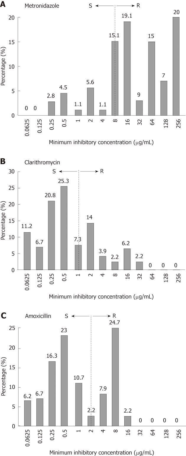
Figure 1 Prevalence of Helicobacter pylori resistance to first-line drugs.
Dotted box represents the distribution of resistant strains at different minimum inhibitory concentrations (MICs). MIC breakpoints were ≥ 8 μg/mL (A), ≥ 1 μg/mL (B) and ≥ 2 μg/mL (C). S: Sensitive; R: Resistant.
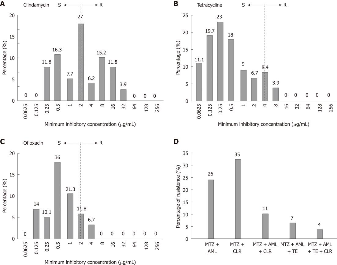
Figure 2 The prevalence of Helicobacter pylori resistance to second-line drugs.
Dotted box represents the distribution of resistant strains at different minimum inhibitory concentrations (MICs). MIC breakpoints were ≥ 1 μg/mL (A), ≥ 4 μg/mL (B) and ≥ 2 μg/mL (C); D: Distribution of multidrug resistant strains. AML: Amoxicillin; CLR: Clarithromycin; MTZ: Metronidazole; TE: Tetracycline. S: Sensitive; R: Resistant.
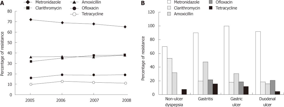
Figure 3 The trend analysis antimicrobial drug resistance in Helicobacter pylori strains (n = 178).
A: From 2005 to 2008, significant increase in clarithromycin resistance was observed by linear regression (P = 0.004); B: Correlation of drug resistant strains in various groups of patients. Significant distribution of metronidazole (P = 0.001), clarithromycin (P < 0.000) and amoxicillin (P = 0.005) resistance was observed by Pearson’s χ2 test. NUD: Non ulcerative dyspepsia; GU: Gastric ulcer; DU: Duodenal ulcer.
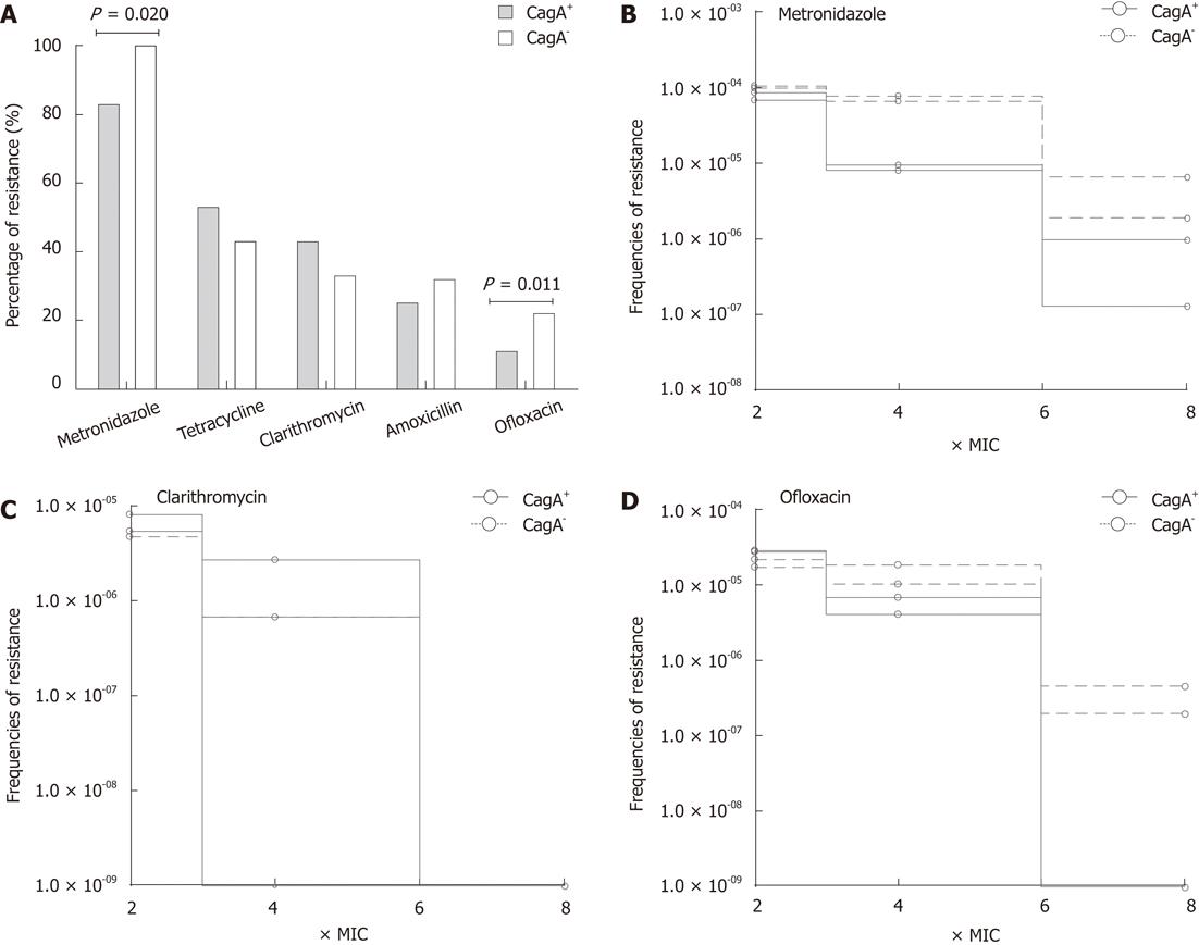
Figure 4 The correlation of antimicrobial drug resistance with cagA gene.
A: Rate of drug resistance in Helicobacter pylori (H. pylori) strains carrying (n = 81) and devoid of (n = 72) of cagA gene. Statistical differences were observed by Pearson’s χ2 test. To determine the effect of cagA carriage on the development of resistance, cagA+ (n = 2) and cagA- (n = 2) H. pylori strains were exposed to the increasing concentrations of metronidazole (B) clarithromycin (C) and ofloxacin (D). Bacterial growth was monitored at different concentration of antibiotic and frequencies of resistant mutants were determined as the colony forming units of H. pylori strain divided by the starting inocula.
-
Citation: Khan A, Farooqui A, Manzoor H, Akhtar SS, Quraishy MS, Kazmi SU. Antibiotic resistance and
cagA gene correlation: A looming crisis ofHelicobacter pylori . World J Gastroenterol 2012; 18(18): 2245-2252 - URL: https://www.wjgnet.com/1007-9327/full/v18/i18/2245.htm
- DOI: https://dx.doi.org/10.3748/wjg.v18.i18.2245


