Copyright
©2012 Baishideng Publishing Group Co.
World J Gastroenterol. May 14, 2012; 18(18): 2188-2196
Published online May 14, 2012. doi: 10.3748/wjg.v18.i18.2188
Published online May 14, 2012. doi: 10.3748/wjg.v18.i18.2188
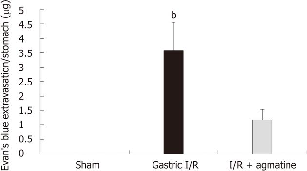
Figure 1 Evan’s blue extravasation as a measure of vascular permeability in the studied groups.
Gastric ischemia reperfusion (I/R) injury induced extravasation of Evan’s blue dye due to increased vascular permeability. Agmatine (AGM) pre-treatment (100 mg/kg, i.p.) significantly attenuated the vascular leakage (gastric I/R vs I/R + agmatine, bP < 0.001). Results are expressed as mean ± SD.
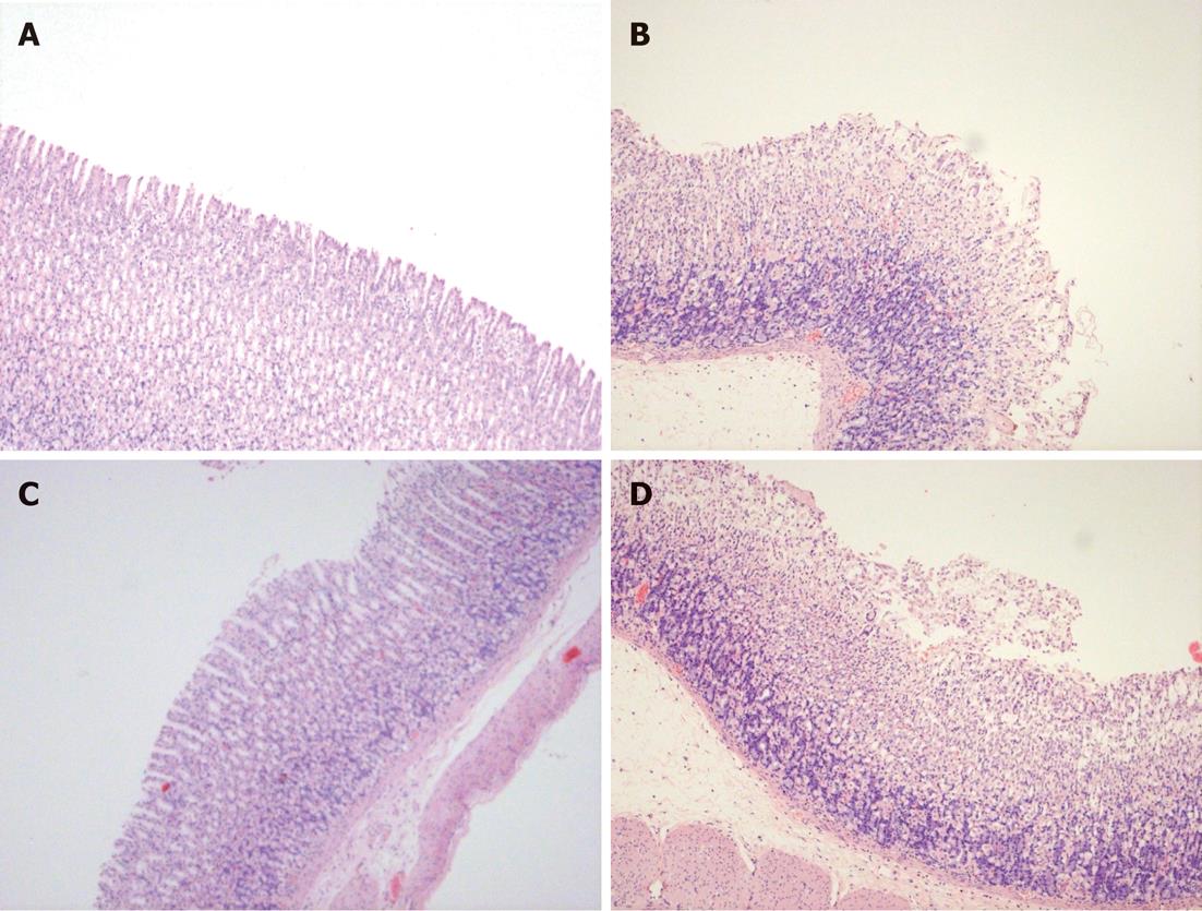
Figure 2 Histological appearance of the gastric mucosa of HE stained sections of the studied groups.
A: Sham-operated showing intact mucosa; B: Gastric ischemia reperfusion (I/R) showing hemorrhages, edema and ulceration; C: Agmatine (AGM) treated group (100 mg/kg, i.p. prior to I/R) with marked preservation of gastric mucosa and disappearance of ulceration and hemorrhages; D: Effect of inhibition of Akt/phosphatidyl inositol-3-kinase (PI3K) (wortmannin 15 μg/kg, i.p.) prior to AGM treatment, showing extensive lesions and salvage of gastric mucosa into the lumen (× 20 magnification).
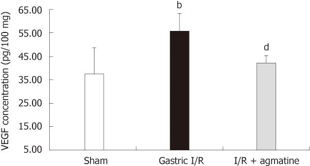
Figure 3 Effect of administration of agmatine on gastric vascular endothelial growth factor tissue level.
Gastric vascular endothelial growth factor (VEGF) protein significantly increased after ischemia reperfusion (I/R) injury, bP < 0.01 vs sham group. Administration of agmatine (100 mg/kg, i.p.) 15 min prior to gastric I/R reduced the VEGF level in gastric tissue homogenate, dP < 0.01 vs I/R group. Results are expressed as mean ± SD.
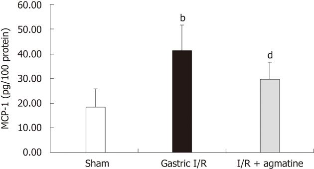
Figure 4 Effect of AGM administration on gastric monocyte chemoattractant protein-1 tissue level.
Gastric monocyte chemoattractant protein-1 (MCP-1) protein significantly increased after ischemia reperfusion (I/R) injury, bP < 0.01 vs sham group. Administration of agmatine (100 mg/kg, i.p.) 15 min prior to gastric I/R reduced the MCP-1 level in gastric tissue homogenate, dP < 0.01 vs I/R group. Results are expressed as mean ± SD.
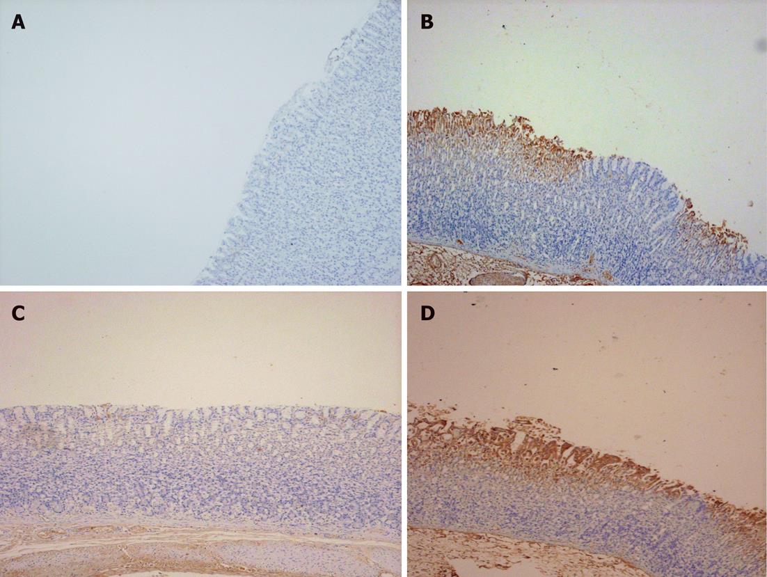
Figure 5 Photomicrographs representative of angiopoietin-1 immunostaining of gastric sections of the studied groups.
A: Sham-operated showing lack of expression of angiopoietin-1 (Ang-1); B: Gastric ischemia reperfusion (I/R) group showing extensive cytoplasmic expression of Ang-1 particularly in areas of congestion and damage; C: Agmatine (AGM) treated group (100 mg/kg, i.p.), with marked attenuation of Ang-1 expression in intact mucosa; D: Wortmannin (15 μg/kg, i.p.) treated group prior to AGM administration abolished the effect of AGM and the expression of Ang-1 is marked (× 20 magnification).
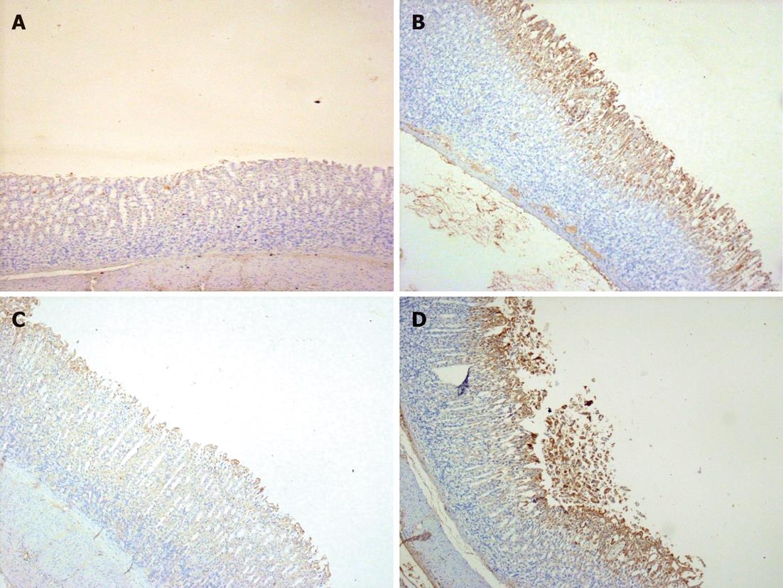
Figure 6 Photomicrographs representative of angiopoietin-2 immunostaining of gastric sections of the studied groups.
A: Sham-operated showing faint expression of angiopoietin-2 (Ang-2); B: Gastric ischemia reperfusion (I/R) group showing extensive cytoplasmic expression of Ang-2 in areas of congestion and damage, and in the submucosa; C: Agmatine (AGM) treated group (100 mg/kg, i.p.), with marked attenuation of Ang-2 expression in intact mucosa; D: Wortmannin (15 μg/kg, i.p.) treated group prior to AGM administration abolished the effect of AGM and the expression of Ang-2 is marked in mucosa and sloughed tissues (× 20 magnification).
- Citation: Masri AAA, Eter EE. Agmatine induces gastric protection against ischemic injury by reducing vascular permeability in rats. World J Gastroenterol 2012; 18(18): 2188-2196
- URL: https://www.wjgnet.com/1007-9327/full/v18/i18/2188.htm
- DOI: https://dx.doi.org/10.3748/wjg.v18.i18.2188


