Copyright
©2012 Baishideng Publishing Group Co.
World J Gastroenterol. Apr 28, 2012; 18(16): 1921-1925
Published online Apr 28, 2012. doi: 10.3748/wjg.v18.i16.1921
Published online Apr 28, 2012. doi: 10.3748/wjg.v18.i16.1921
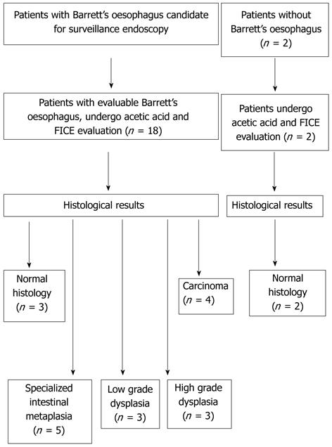
Figure 1 Patient selection for the present study.
FICE: Fujinon intelligent chromoendoscopy.
Figure 2 Acetic acid and Fujinon intelligent chromoendoscopy image of the oesophagus.
Specialized intestinal metaplasia using high definition white light (A), 2% acetic acid (B), and the combination of acetic acid with Fujinon intelligent chromoendoscopy (FICE) 4 (C), FICE 0 (D) and FICE 7 (E).
Figure 3 Acetic acid and Fujinon intelligent chromoendoscopy image of an oesophageal carcinoma.
Irregular pit pattern and abnormal vascularisation is shown with 2% acetic acid (A), or following the combination of acetic acid and Fujinon intelligent chromoendoscopy (FICE) 4 (B), FICE 0 (C) and FICE 7 (D).
- Citation: Camus M, Coriat R, Leblanc S, Brezault C, Terris B, Pommaret E, Gaudric M, Chryssostalis A, Prat F, Chaussade S. Helpfulness of the combination of acetic acid and FICE in the detection of Barrett's epithelium and Barrett's associated neoplasias. World J Gastroenterol 2012; 18(16): 1921-1925
- URL: https://www.wjgnet.com/1007-9327/full/v18/i16/1921.htm
- DOI: https://dx.doi.org/10.3748/wjg.v18.i16.1921


