Copyright
©2012 Baishideng Publishing Group Co.
World J Gastroenterol. Apr 28, 2012; 18(16): 1915-1920
Published online Apr 28, 2012. doi: 10.3748/wjg.v18.i16.1915
Published online Apr 28, 2012. doi: 10.3748/wjg.v18.i16.1915
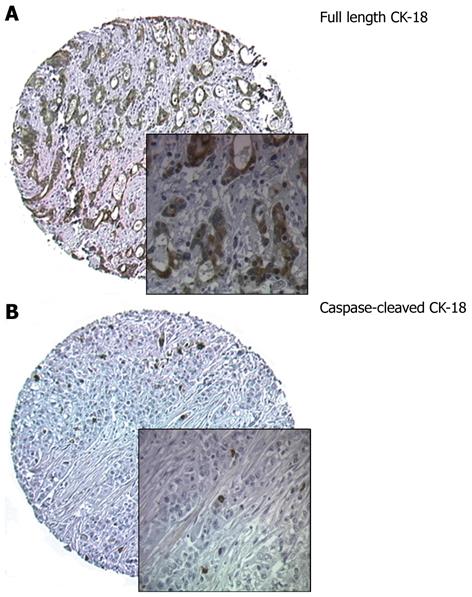
Figure 1 Immunohistochemical staining of full length cytokeratin-18 and caspase-cleaved cytokeratin-18.
A: Immunohistochemical staining of full length cytokeratin-18 (CK-18) showing strong cytoplasmic staining; B: Immunohistochemical staining for caspase-cleaved CK-18. Cores from tumour showing positively-stained apoptotic cells. Original magnification × 100; insets × 400.
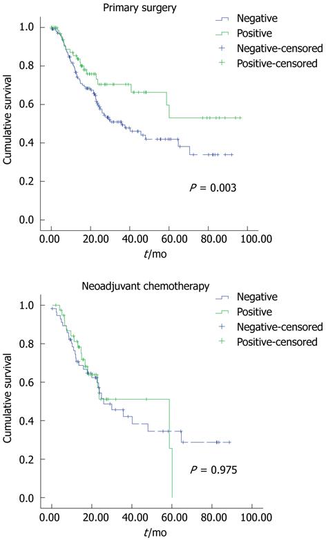
Figure 2 Kaplan Meier curves representing the relationship between caspase-cleaved cytokeratin-18 and disease-specific survival.
A: In months from time of diagnosis in patients who received primary surgery only; B: In patients who received neoadjuvant chemotherapy.
- Citation: Fareed KR, Soomro IN, Hameed K, Arora A, Lobo DN, Parsons SL, Madhusudan S. Caspase-cleaved cytokeratin-18 and tumour regression in gastro-oesophageal adenocarcinomas treated with neoadjuvant chemotherapy. World J Gastroenterol 2012; 18(16): 1915-1920
- URL: https://www.wjgnet.com/1007-9327/full/v18/i16/1915.htm
- DOI: https://dx.doi.org/10.3748/wjg.v18.i16.1915


