Copyright
©2012 Baishideng Publishing Group Co.
World J Gastroenterol. Apr 21, 2012; 18(15): 1840-1844
Published online Apr 21, 2012. doi: 10.3748/wjg.v18.i15.1840
Published online Apr 21, 2012. doi: 10.3748/wjg.v18.i15.1840
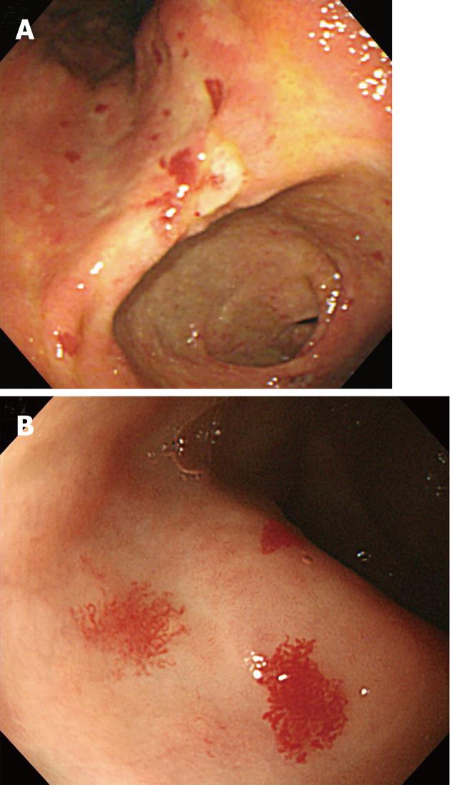
Figure 1 Multiple angiodysplastic lesions were noted in the entire stomach upon endoscopic examination.
A: A view of the lesser curvature of the angle showing multiple scattered angiodysplastic lesions; B: A view of the anterior wall of the antrum, magnified view of angiodysplastic lesions showing vessel-pulled appearance.
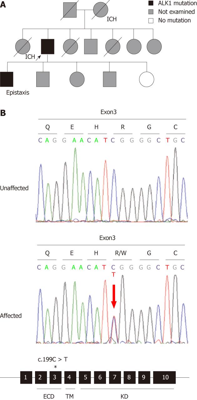
Figure 2 Pedigree and genetic analysis of an hereditary hemorrhagic telangiectasia family.
A: Pedigree of a family with genetic mutations and/or symptoms of hereditary hemorrhagic telangiectasia (HHT) and intracranial hemorrhage (ICH). The proband is indicated by an arrow. A divided symbol represents the individual with ICH. Deceased individuals are indicated by a slash; B: Genetic studies of unaffected and affected family members. The affected member had a heterozygous activin receptor-like kinase 1 (ALK1) mutation (c.199C > T; p.Arg67Trp). The amino acid translation is shown above each codon. The mutation was found in exon 3, indicated by an asterisk; 3. Protein domains of ALK1 are indicated under the exons: extracellular domain (ECD), transmembrane domain (TM), and kinase domain (KD).
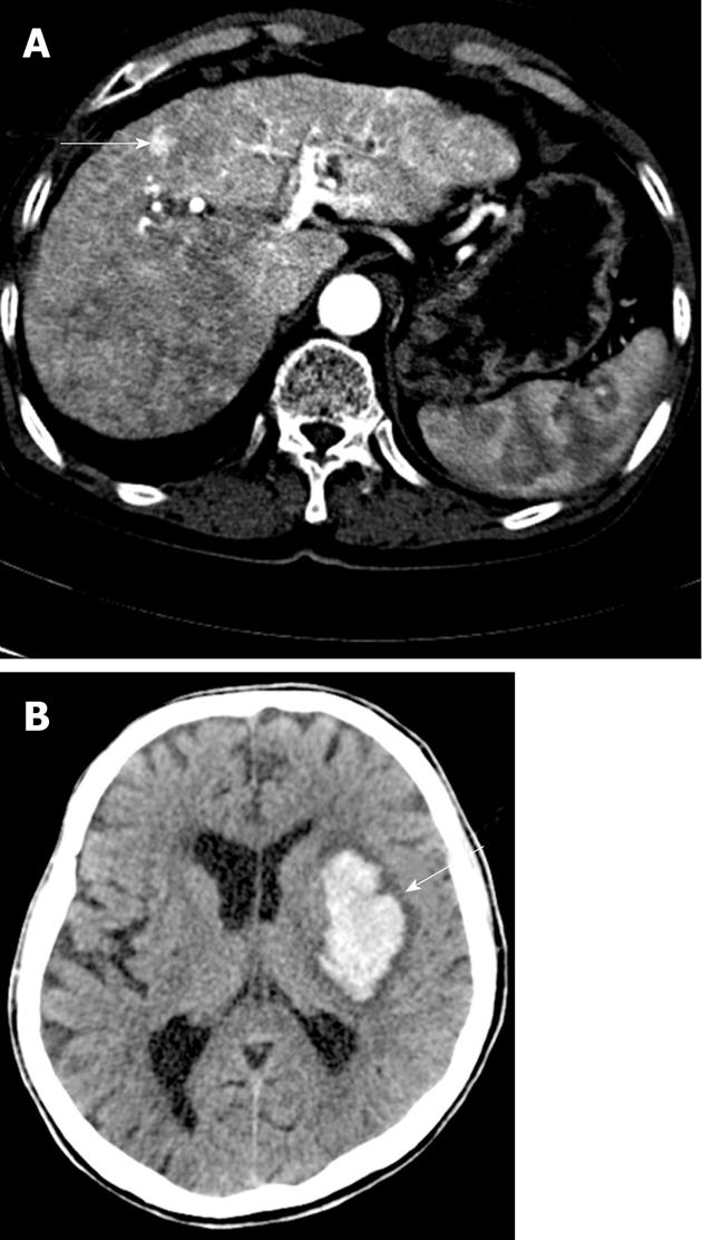
Figure 3 Hepatic arterio-venous shunt and cerebral hemorrhage.
A: Abdominal computed tomography (CT) showing an intra-hepatic arterio-venous shunt; B: Cerebral hemorrhage was noted in the left basal ganglia on brain CT.
- Citation: Ha M, Kim YJ, Kwon KA, Hahm KB, Kim MJ, Kim DK, Lee YJ, Oh SP. Gastric angiodysplasia in a hereditary hemorrhagic telangiectasia type 2 patient. World J Gastroenterol 2012; 18(15): 1840-1844
- URL: https://www.wjgnet.com/1007-9327/full/v18/i15/1840.htm
- DOI: https://dx.doi.org/10.3748/wjg.v18.i15.1840


