Copyright
©2012 Baishideng Publishing Group Co.
World J Gastroenterol. Apr 7, 2012; 18(13): 1545-1551
Published online Apr 7, 2012. doi: 10.3748/wjg.v18.i13.1545
Published online Apr 7, 2012. doi: 10.3748/wjg.v18.i13.1545
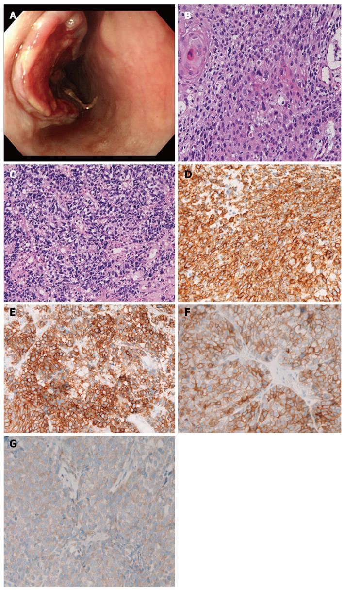
Figure 1 Case 1.
A: Endoscopy. An ulcerated tumor is seen in the esophagus; B: Histology of the squamous cell carcinoma element of the esophageal tumor. Keratinization is seen [hematoxylin and eosin (HE), x 200]; C: Small cell carcinoma element of the esophageal carcinoma. The tumor cells show characteristic morphologies of small cell carcinoma (HE, x 200); D: Cytokeratins are expressed in the small cell carcinoma component (x 200); E: Synaptophysin is expressed in the small cell carcinoma component (x 200); F: KIT is expressed in the small cell carcinoma component (x 200); G: Platelet-derived growth factor-α is expressed in the small cell carcinoma component (x 200).
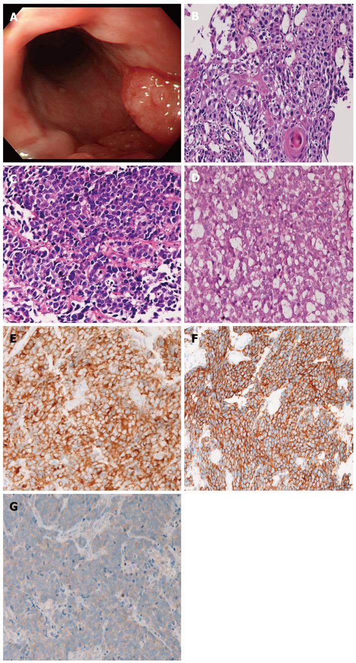
Figure 2 Case 2.
A: Endoscopy. An elevated tumor is seen in the esophagus; B: Histology of the squamous cell carcinoma element of the esophageal tumor. A cancer pearl is seen [hematoxylin and eosin (HE), x 200]; C: Small cell carcinoma element of the esophageal carcinoma. The tumor cells show characteristic morphologies of small cell carcinoma (HE, x 200); D: Adenocarcinomatous element shows focal tubular formations (HE, x 200); E: CD56 is expressed in the small cell carcinoma component (x 200); F: KIT is expressed in the small cell carcinoma component (x 200); G: Platelet-derived growth factor-α is expressed in the small cell carcinoma component (x 200).
- Citation: Terada T, Maruo H. Esophageal combined carcinomas: Immunohoistochemical and molecular genetic studies. World J Gastroenterol 2012; 18(13): 1545-1551
- URL: https://www.wjgnet.com/1007-9327/full/v18/i13/1545.htm
- DOI: https://dx.doi.org/10.3748/wjg.v18.i13.1545


