Copyright
©2012 Baishideng Publishing Group Co.
World J Gastroenterol. Jan 7, 2012; 18(1): 1-15
Published online Jan 7, 2012. doi: 10.3748/wjg.v18.i1.1
Published online Jan 7, 2012. doi: 10.3748/wjg.v18.i1.1
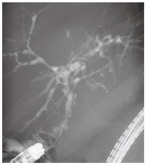
Figure 1 Endoscopic retrograde cholangiogram in a patient with recurrent primary sclerosing cholangitis, showing multifocal stenosis with intervening saccular dilatations affecting both intrahepatic and extrahepatic bile ducts.
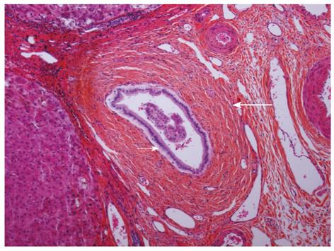
Figure 2 Histological changes demonstrated in a biopsy from a patient with recurrent primary sclerosing cholangitis.
Bile duct (small arrow) surrounded by collar of connective tissue with concentric layers of collagen fibers (large arrow) illustrating the typical periductal lamellar fibrosis. (Original magnification, x 100).
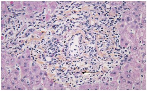
Figure 3 Histological changes seen in a biopsy with acute cellular rejection in a patient with primary sclerosing cholangitis.
Portal tract with mixed inflammatory infiltrate containing blastic lymphocytes and eosinophils. Subendothelial localization of the inflammatory cells in a portal vein branch (small arrow). Inflammation of small bile duct (large arrow). (Original magnification, x 400).
- Citation: Fosby B, Karlsen TH, Melum E. Recurrence and rejection in liver transplantation for primary sclerosing cholangitis. World J Gastroenterol 2012; 18(1): 1-15
- URL: https://www.wjgnet.com/1007-9327/full/v18/i1/1.htm
- DOI: https://dx.doi.org/10.3748/wjg.v18.i1.1


