Copyright
©2011 Baishideng Publishing Group Co.
World J Gastroenterol. Mar 7, 2011; 17(9): 1167-1173
Published online Mar 7, 2011. doi: 10.3748/wjg.v17.i9.1167
Published online Mar 7, 2011. doi: 10.3748/wjg.v17.i9.1167
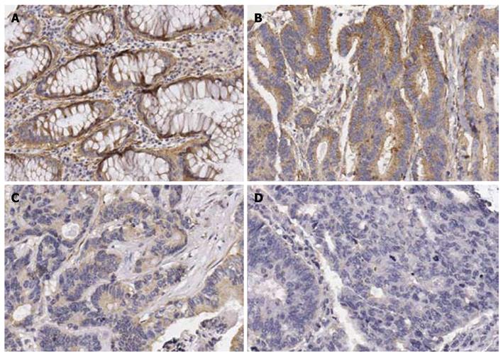
Figure 1 Immunohistochemical staining of interleukin-24 in rectal tissue (200 ×).
A: In the normal rectal mucosa tissue, the non-neoplastic glandular epithelial cells were strongly positive for IL-24; B: In rectal cancer, a well-differentiated adenocarcinoma showed strong positive expression of IL-24; C: A moderately differentiated adenocarcinoma showed weak positive expression of IL-24; D: A poorly differentiated adenocarcinoma showed negative expression of IL-24. IL-24: Interleukin-24.
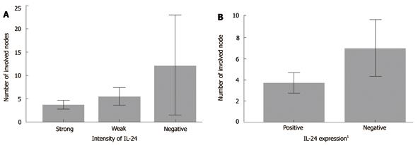
Figure 2 The number of involved lymph nodes according to interleukin-24 intensity (A) and expression status (B) in node-positive rectal cancer patients.
1Negative (n = 9) and weak (n = 31) intensities were classified as negative expression, and strong intensity (n = 50) as positive. IL: Interleukin.
- Citation: Choi Y, Roh MS, Hong YS, Lee HS, Hur WJ. Interleukin-24 is correlated with differentiation and lymph node numbers in rectal cancer. World J Gastroenterol 2011; 17(9): 1167-1173
- URL: https://www.wjgnet.com/1007-9327/full/v17/i9/1167.htm
- DOI: https://dx.doi.org/10.3748/wjg.v17.i9.1167


