Copyright
©2011 Baishideng Publishing Group Co.
World J Gastroenterol. Feb 14, 2011; 17(6): 717-726
Published online Feb 14, 2011. doi: 10.3748/wjg.v17.i6.717
Published online Feb 14, 2011. doi: 10.3748/wjg.v17.i6.717
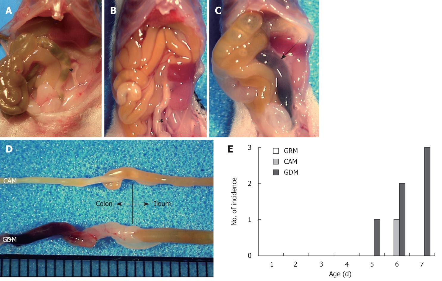
Figure 1 Macroscopic views of milk-fed mice with colonic hemorrhage.
Representative macroscopic views of newborn mice that were fed with glutamine-rich milk (GRM) (A), complete amino acid milk (CAM) (B), and glutamine-deleted milk (GDM) (C) are shown. Compared to the colons of GRM-mice and CAM-mice (asterisks), those of the GDM-mice appeared distended and edematous, with a pool of blood (arrow); D: Close-up views of the resected intestines from a CAM-mouse and a GDM-mouse, shown for comparison; E: Bar chart of the number of mice with melena on each day.
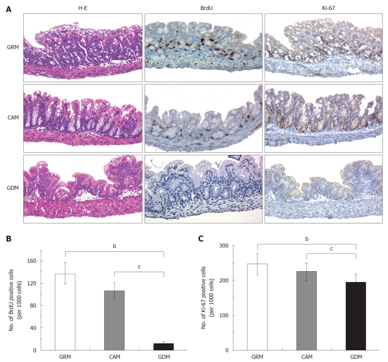
Figure 2 Glutamine depletion induces severe damage of the colonic epithelial structure with reduced epithelial cell growth.
A: Representative pictures of H-E staining (left panels) and immunohistochemical staining for BrdU (middle panels) and Ki-67 (right panels). Each colonic epithelium was taken from infant mice fed with glutamine-rich milk (GRM), complete amino acid milk (CAM), or glutamine-deleted milk (GDM). Magnification, × 200. Positive stained epithelial cells with BrdU (B) and Ki-67 (C) were counted and compared with each group in a histogram. bP≤ 0.001; cP < 0.05.
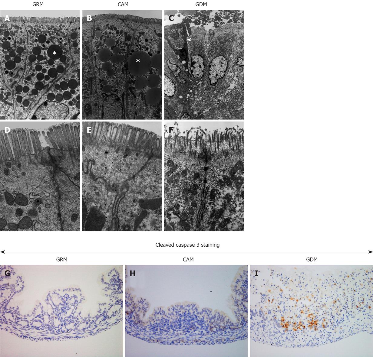
Figure 3 Apoptotic changes observed in the damaged colonic epithelium.
Electron micrographs of colonic epithelia obtained from infant mice with glutamine-rich milk (GRM) (A, D), complete amino acid milk (CAM) (B, E), and glutamine-deleted milk (GDM) (C, F) at low magnification (A-C) and high magnification (D-F). Asterisks (*) represent lipid droplets and the arrow shows an apoptotic cell. G-I: Optical microscopic views (magnification, × 200) of immunohistochemistry for cleaved caspase-3 as a marker of apoptosis using resected tissues from the same mice (G: GRM, H: CAM, I: GDM).
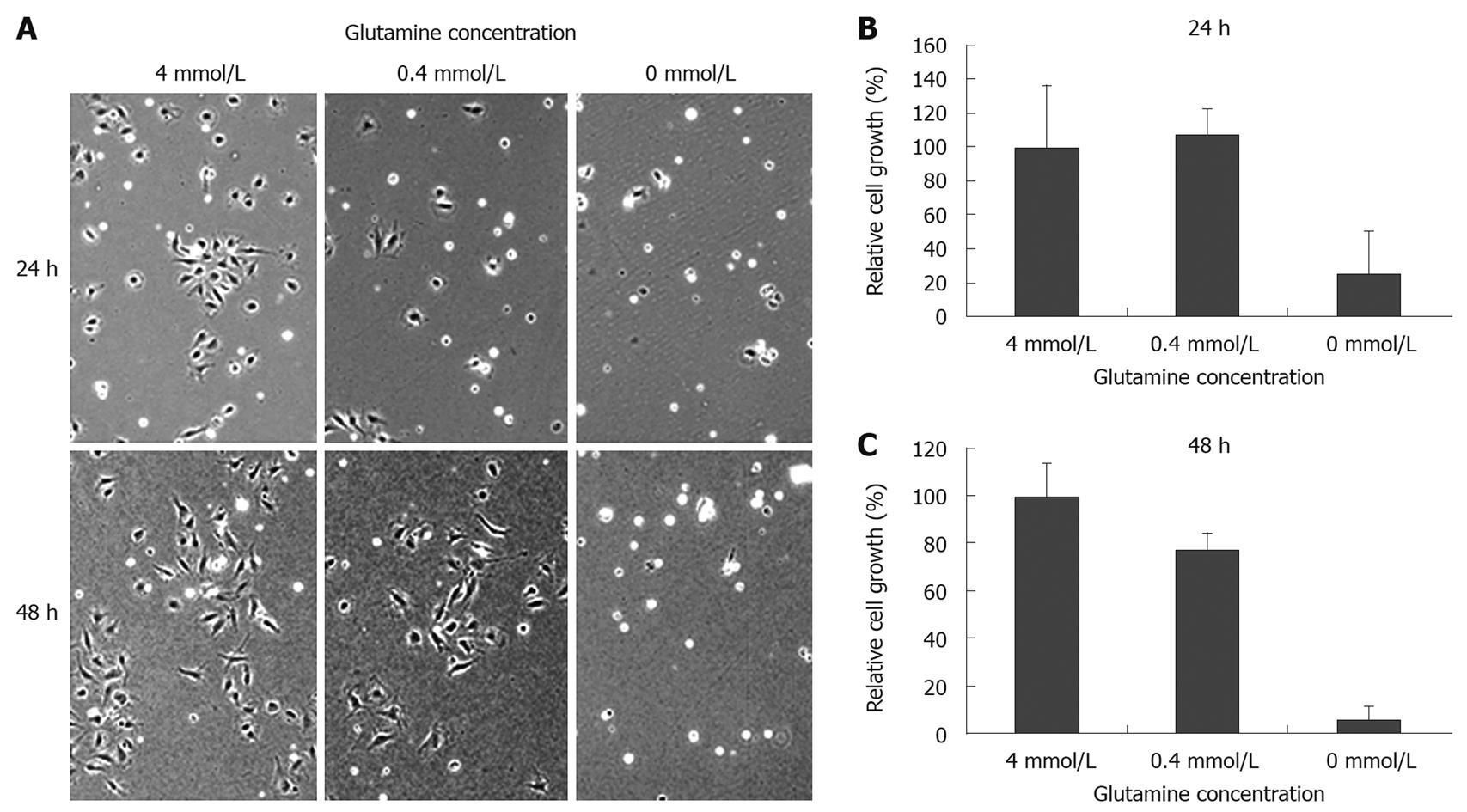
Figure 4 Glutamine depletion suppresses cell proliferation of cultured intestinal epithelial cells.
A: IEC6 rat intestinal epithelial cells were treated with media containing different amounts of glutamine (0, 0.4 and 4 mmol/L); Cell morphology and cell number were observed at the indicated time points (B: 24 h, C: 48 h).
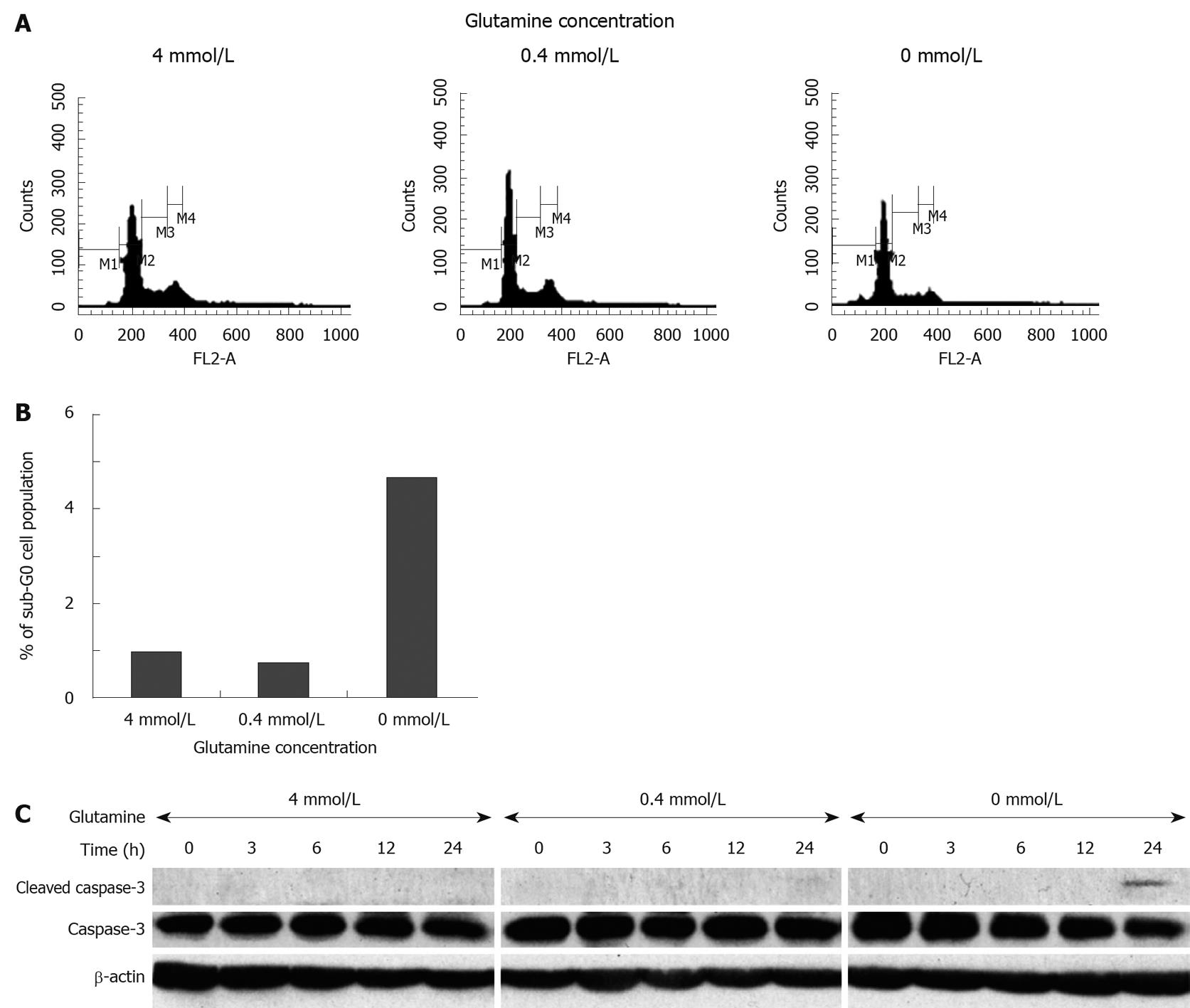
Figure 5 Potent antiproliferative effect of glutamine depletion results in increased cleavage of caspase-3.
Cell cycle distribution under culture condition with different concentrations of glutamine was analyzed by flow cytometry (A) and cell populations at the sub-G0 phase were compared with each other (B); C: Immunoblotting for caspase-3 and cleaved caspase-3 revealed the induction of apoptosis in IEC6 cells after 24 h of culture without glutamine. Each experiment was independently repeated three times and the representative data among the similar results are shown.
- Citation: Motoki T, Naomoto Y, Hoshiba J, Shirakawa Y, Yamatsuji T, Matsuoka J, Takaoka M, Tomono Y, Fujiwara Y, Tsuchita H, Gunduz M, Nagatsuka H, Tanaka N, Fujiwara T. Glutamine depletion induces murine neonatal melena with increased apoptosis of the intestinal epithelium. World J Gastroenterol 2011; 17(6): 717-726
- URL: https://www.wjgnet.com/1007-9327/full/v17/i6/717.htm
- DOI: https://dx.doi.org/10.3748/wjg.v17.i6.717


