Copyright
©2011 Baishideng Publishing Group Co.
World J Gastroenterol. Feb 7, 2011; 17(5): 677-680
Published online Feb 7, 2011. doi: 10.3748/wjg.v17.i5.677
Published online Feb 7, 2011. doi: 10.3748/wjg.v17.i5.677
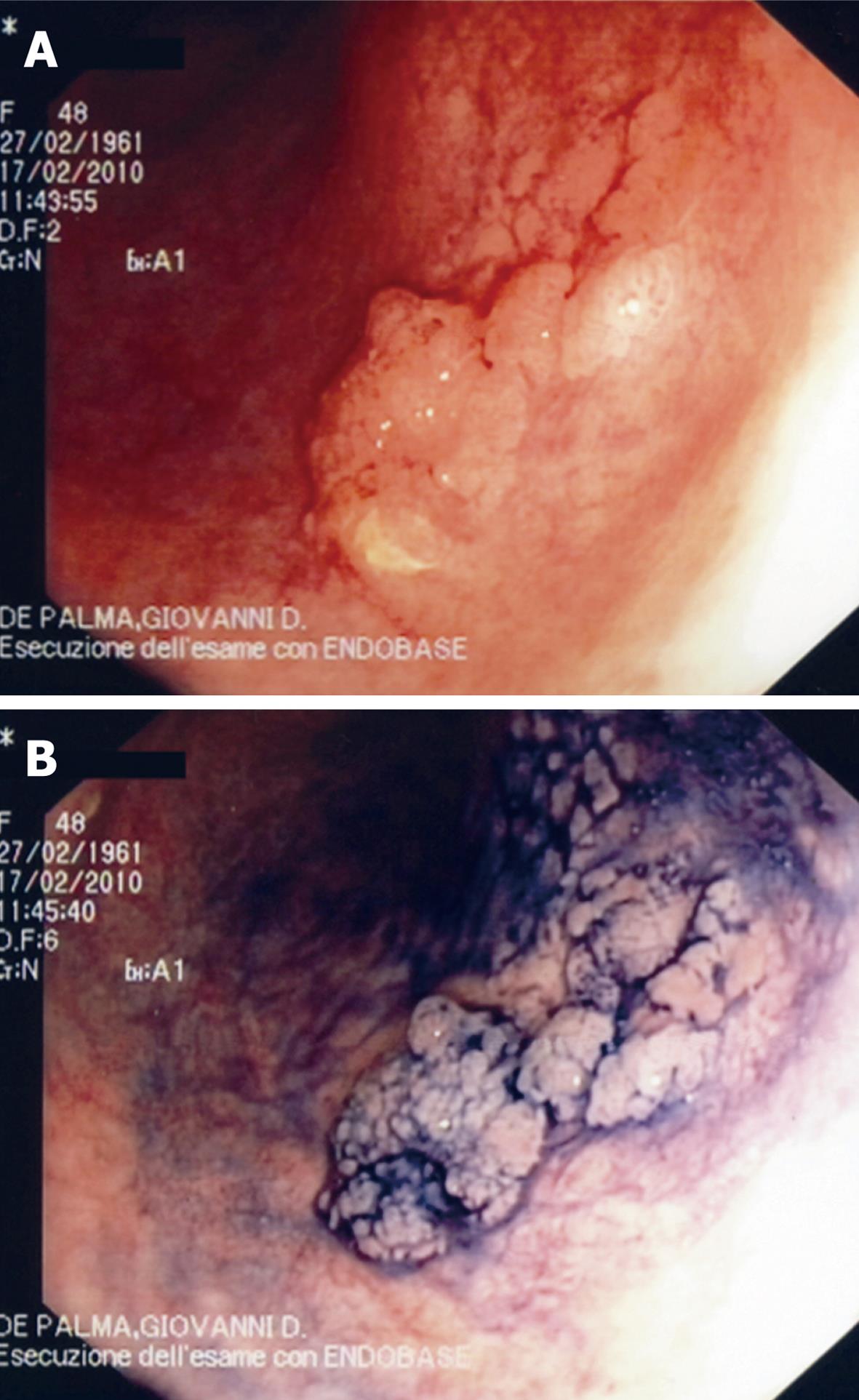
Figure 1 Conventional “white light” imaging of a plaque-like lesion of sigmoid colon in ulcerative colitis (A) and 0.
5% indigo carmine chromoscopy of the lesion (B). The lesion is “unmasked” and clearly delineated.
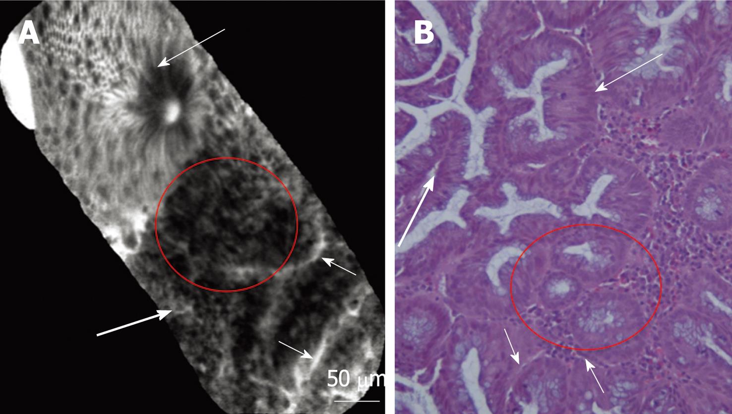
Figure 2 Confocal (A) and histological (B) images of colonic mucosa showing the switch from normal mucosa to inflamed mucosa.
Normal crypt architecture is classically represented by ordered and regular crypt orifices covered by a homogeneous epithelial layer with visible “black-hole” goblet cells within the subcellular matrix (long thin arrows). Inflamed mucosa showing irregular arrangement of crypts, crypt fusion (red circles) and capillaries alterations (short arrows) and inflammatory cells (lymphocytes: long thick arrows). Magnification, × 200.
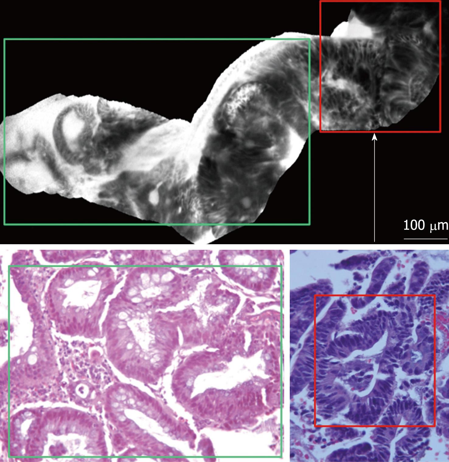
Figure 3 Confocal images of colonic mucosa evidencing the switch from the inflamed mucosa, to the neoplastic mucosa.
Inflamed mucosa (green rectangle) is characterized by dilation of crypt openings, enlarged spaces between crypt, and microvascular alterations with fluorescein leaks into the crypt lumen (white arrow) therefore making the lumen brighter than the surrounding epithelium. Dysplastic mucosa (red rectangle) is characterized by “dark” cells, irregular architectural patterns with villiform structures and a “dark” epithelial border. Histology images show high-power hematoxylin and eosin stain of the tissue sampled, evidencing respectively inflamed area with features suggestive of chronic ulcerative colitis (green rectangle) and low grade dysplasia (red rectangle). Magnification, × 200.
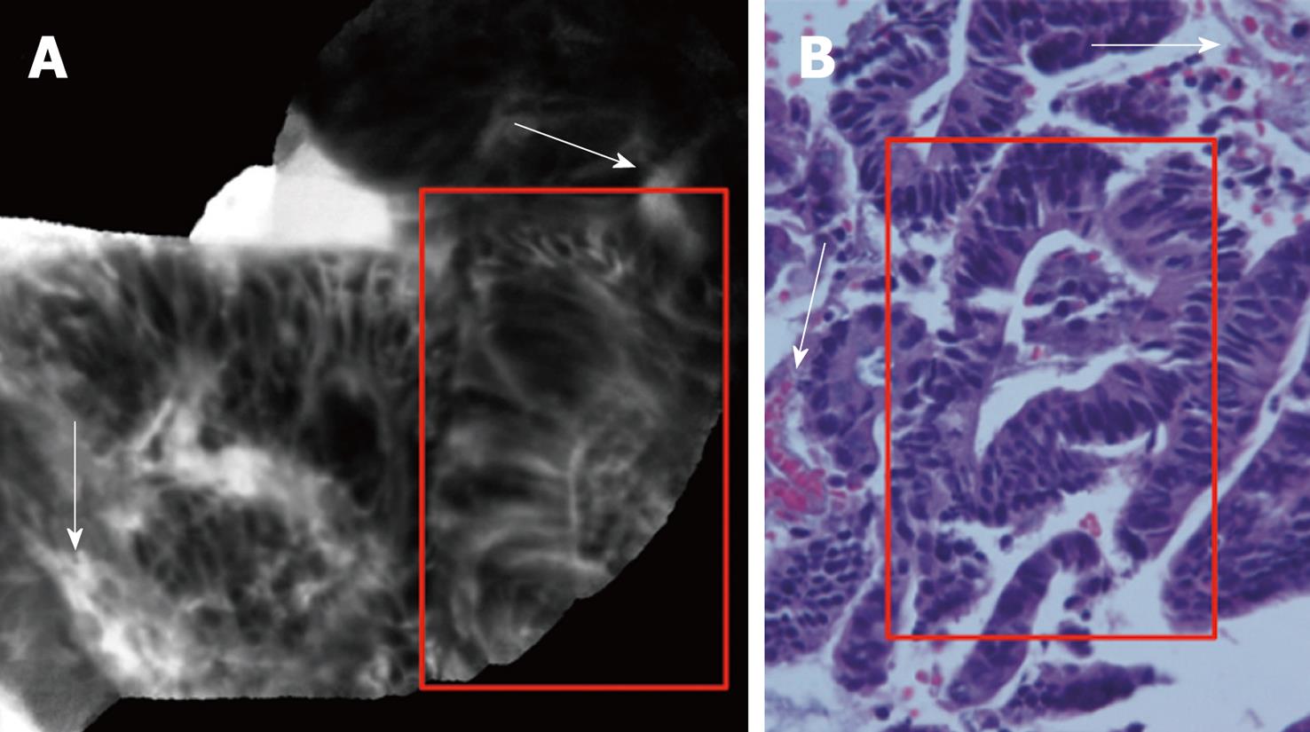
Figure 4 Confocal (A) and histological (B) images of dysplasia-associated lesional mass showing “dark” cells, with mucin depletion and goblet cell/crypt density attenuation; the architectural pattern is irregular, as well as the epithelial thickness, with villiform structures and “dark” epithelial border (red rectangles).
There is gross distortion of the vascular architecture with tortuous and dilated vessels (white arrows). The hematoxylin and eosin stain histology shows a low grade dysplasia (red rectangle; hematoxylin and eosin staining; original magnification, × 200).
- Citation: Palma GDD, Staibano S, Siciliano S, Maione F, Siano M, Esposito D, Persico G. In vivo characterization of DALM in ulcerative colitis with high-resolution probe-based confocal laser endomicroscopy. World J Gastroenterol 2011; 17(5): 677-680
- URL: https://www.wjgnet.com/1007-9327/full/v17/i5/677.htm
- DOI: https://dx.doi.org/10.3748/wjg.v17.i5.677


