Copyright
©2011 Baishideng Publishing Group Co.
World J Gastroenterol. Dec 28, 2011; 17(48): 5324-5326
Published online Dec 28, 2011. doi: 10.3748/wjg.v17.i48.5324
Published online Dec 28, 2011. doi: 10.3748/wjg.v17.i48.5324
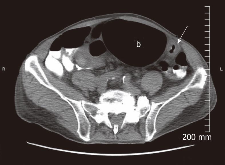
Figure 1 Computed tomography of the abdomen demonstrating circumferential thickening of a long segment of the distal colon (marked with white arrow) with a proximal dilated segment (marked with “b” inside the lumen of the dilated segment).
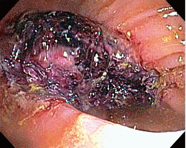
Figure 2 Colonoscopy image demonstrating friable mass occupying up to half the circumference of the splenic flexure.
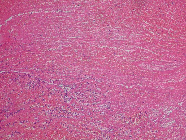
Figure 3 Hematoxylin and eosin high power microscopic image demonstrating acute colitis with marked fibrinopurulent exudates.
- Citation: Deepak P, Devi R. Ischemic colitis masquerading as colonic tumor: Case report with review of literature. World J Gastroenterol 2011; 17(48): 5324-5326
- URL: https://www.wjgnet.com/1007-9327/full/v17/i48/5324.htm
- DOI: https://dx.doi.org/10.3748/wjg.v17.i48.5324


