Copyright
©2011 Baishideng Publishing Group Co.
World J Gastroenterol. Dec 14, 2011; 17(46): 5110-5116
Published online Dec 14, 2011. doi: 10.3748/wjg.v17.i46.5110
Published online Dec 14, 2011. doi: 10.3748/wjg.v17.i46.5110
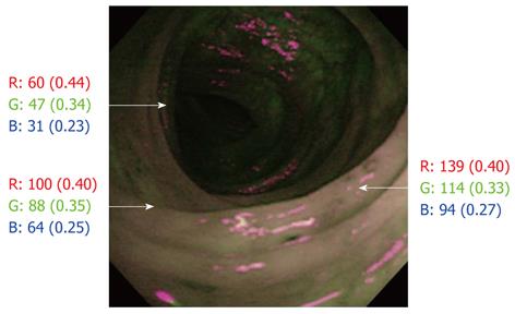
Figure 1 Endoscopic photograph by autofluorescence imaging showing the actual counts of red, green and blue colors based on the RGB additive model in different mucosal areas.
The actual counts of red, green and blue colors varied depending on the brightness of the examined area, but the abundance of the actual counts, in parentheses, was approximately constant in the same picture.
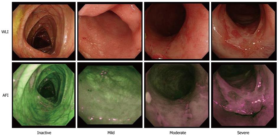
Figure 2 Representative endoscopic photographs of ulcerative colitis using white light imaging (upper row) and autofluorescence imaging (lower row) at the same sites according to the level of endoscopic ulcerative colitis activity (inactive, mild, moderate and severe).
The color of the large intestinal mucosa on the autofluorescence imaging (AFI) changes by degrees, from green to grayish and magenta color. WLI: White light imaging.
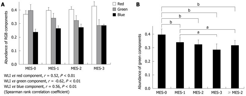
Figure 3 Comparison of overall mucosal inflammation on white light imaging and the abundance of RGB color components on autofluorescence imaging.
A: The green color component corresponds most often with sites of mucosal inflammation in different colonoscopy images (according to Spearman rank correlation coefficient); B: There are significant differences in the green color component values between Mayo endoscopic subscore (MES)-0 and MES-1 (bP < 0.01), between MES-0 and MES-2 (bP < 0.01), between MES-0 and MES-3 (bP < 0.01), between MES-1 and MES-3 (aP < 0.05), between MES-0 and ≥ MES-2 (bP < 0.01) and between MES-1 and ≥ MES-2 (aP < 0.05), but not between MES-1 and MES-2 or between MES-2 and MES-3 (Tukey-Kramer multiple comparison test). WLI: White light imaging.
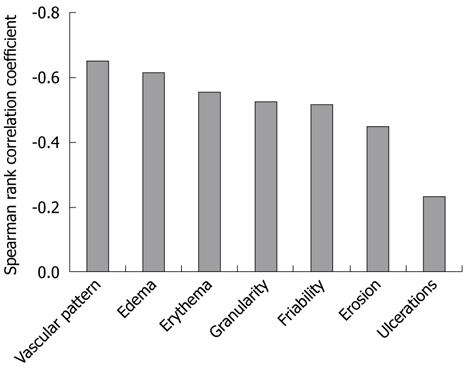
Figure 4 Comparison of seven endoscopic ulcerative colitis features on white light imaging and the green color component on autofluorescence imaging.
The green color component on autofluorescence imaging is well correlated with vascular pattern (r = -0.65, P < 0.01) and edema (r = -0.62, P < 0.01) scores on WLI, but not with the ulcer score (r = -0.23, P < 0.01) (Spearman rank correlation coefficient). WLI: White light imaging.
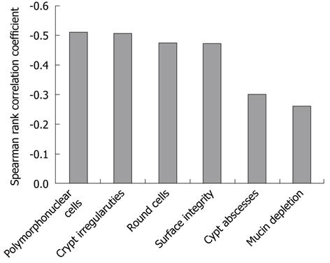
Figure 5 Comparison of histology scores, six histological characteristic ulcerative colitis features and the green color component on autofluorescence imaging.
The autofluorescence imaging green color component is relatively well correlated with the scores for polymorphonuclear cells in the lamina propria (r = -0.51, P < 0.01) and crypt architectural irregularities (r = -0.51, P < 0.01), but not with the scores for crypt abscesses (r = -0.30, P < 0.01), and mucin depletion (r = -0.26, P < 0.01) based on Spearman rank correlation coefficient.
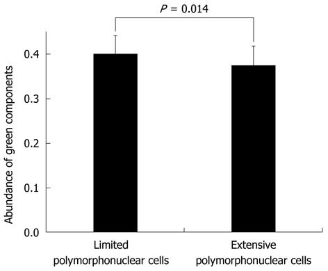
Figure 6 Comparison of limited (combined grades 0 and 1) and extensive (combined grades 2 and 3) polymorphonuclear cell infiltration of the green color components in Mayo endoscopic subscore-0.
The mean number of green color components for images classified into the amount of polymorphonuclear cell infiltration in Mayo endoscopic subscore (MES)-0 is 0.399 ± 0.042 for limited and 0.375 ± 0.044 for extensive infiltration. There are significant differences in green color component values between limited and extensive polymorphonuclear cell infiltration in MES-0 (P = 0.014, by Student’s t-test).
- Citation: Osada T, Arakawa A, Sakamoto N, Ueyama H, Shibuya T, Ogihara T, Yao T, Watanabe S. Autofluorescence imaging endoscopy for identification and assessment of inflammatory ulcerative colitis. World J Gastroenterol 2011; 17(46): 5110-5116
- URL: https://www.wjgnet.com/1007-9327/full/v17/i46/5110.htm
- DOI: https://dx.doi.org/10.3748/wjg.v17.i46.5110


