Copyright
©2011 Baishideng Publishing Group Co.
World J Gastroenterol. Nov 28, 2011; 17(44): 4932-4936
Published online Nov 28, 2011. doi: 10.3748/wjg.v17.i44.4932
Published online Nov 28, 2011. doi: 10.3748/wjg.v17.i44.4932
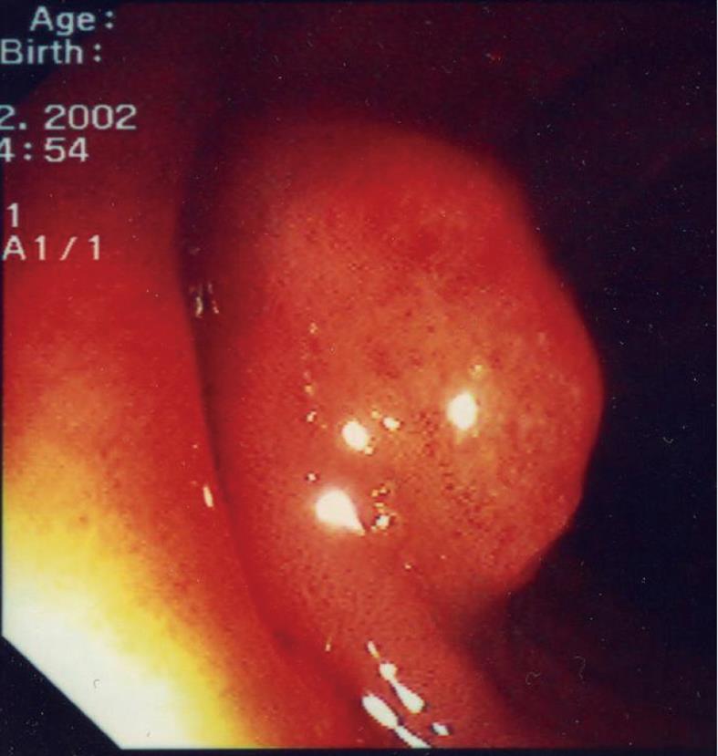
Figure 1 This photograph shows a typical endoscopic appearance of a larger cysts with a reddened overlying mucosa.
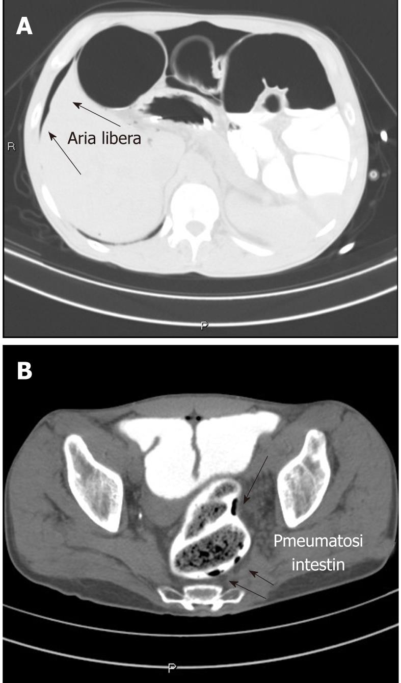
Figure 2 Computed tomography scan image.
A: Presence of free air (arrows) in the abdomen; B: Presence of air in the bowel wall (arrows).
- Citation: Azzaroli F, Turco L, Ceroni L, Galloni SS, Buonfiglioli F, Calvanese C, Mazzella G. Pneumatosis cystoides intestinalis. World J Gastroenterol 2011; 17(44): 4932-4936
- URL: https://www.wjgnet.com/1007-9327/full/v17/i44/4932.htm
- DOI: https://dx.doi.org/10.3748/wjg.v17.i44.4932


