Copyright
©2011 Baishideng Publishing Group Co.
World J Gastroenterol. Nov 21, 2011; 17(43): 4779-4786
Published online Nov 21, 2011. doi: 10.3748/wjg.v17.i43.4779
Published online Nov 21, 2011. doi: 10.3748/wjg.v17.i43.4779
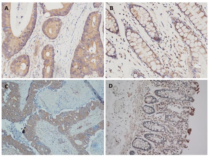
Figure 1 Immunohistochemical photomicrographs of Beclin-1 and LC-3 in colorectal cancer tissues and colorectal normal tissues (200 ×).
A, B: Beclin-1 displayed strongest cytoplasm staining in colorectal cancer (CRC) tissues and weak positive staining in normal tissues: C, D: LC3 displayed intense granular staining of the cytoplasm in CRC tissues but absent in normal tissues.
Figure 2 Progression-free survival of advanced colorectal cancer patients with low and high Beclin-1 expression in cetuximab-containing chemotherapy group.
The median PFS of the patients with low and high Beclin-1 expression (a low expression defined as immuhistochemistry score < 6.0 and a high expression defined as immuhistochemistry score ≥ 6.0.) was 9.0 mo and 3.0 mo respectively (P = 0.01). PFS: Progression-free survival.
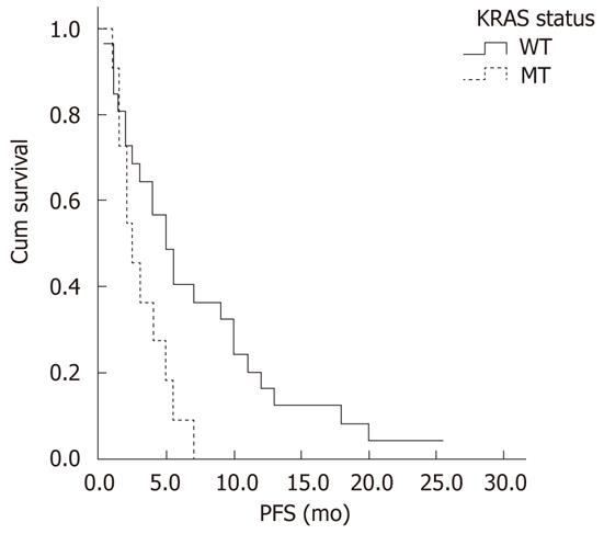
Figure 3 Progression-free survival of advanced colorectal cancer patients with KRAS wild type and patients with KRAS mutation treated by cetuximab-containing chemotherapy.
Thirty-seven of 45 patients were successfully tested KRAS gene in cetuximab-containing chemotherapy group, the median PFS of patients with KRAS wild type was significantly higher than that of patients with KRAS mutation ( 5.0 mo and 2.5 mo, respectively, P = 0.02). PFS: Progression-free survival.
- Citation: Guo GF, Jiang WQ, Zhang B, Cai YC, Xu RH, Chen XX, Wang F, Xia LP. Autophagy-related proteins Beclin-1 and LC3 predict cetuximab efficacy in advanced colorectal cancer. World J Gastroenterol 2011; 17(43): 4779-4786
- URL: https://www.wjgnet.com/1007-9327/full/v17/i43/4779.htm
- DOI: https://dx.doi.org/10.3748/wjg.v17.i43.4779


