Copyright
©2011 Baishideng Publishing Group Co.
World J Gastroenterol. Oct 28, 2011; 17(40): 4539-4541
Published online Oct 28, 2011. doi: 10.3748/wjg.v17.i40.4539
Published online Oct 28, 2011. doi: 10.3748/wjg.v17.i40.4539
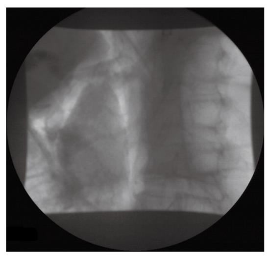
Figure 1 Free gas in the retroperitoneal space.
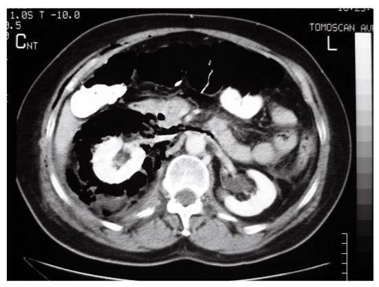
Figure 2 Computed tomography scan showing the presence of air in the retroperitoneal space and subcutaneous emphysema.
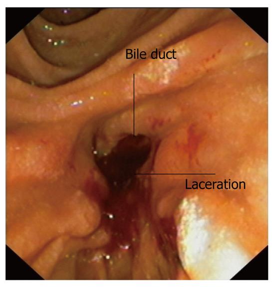
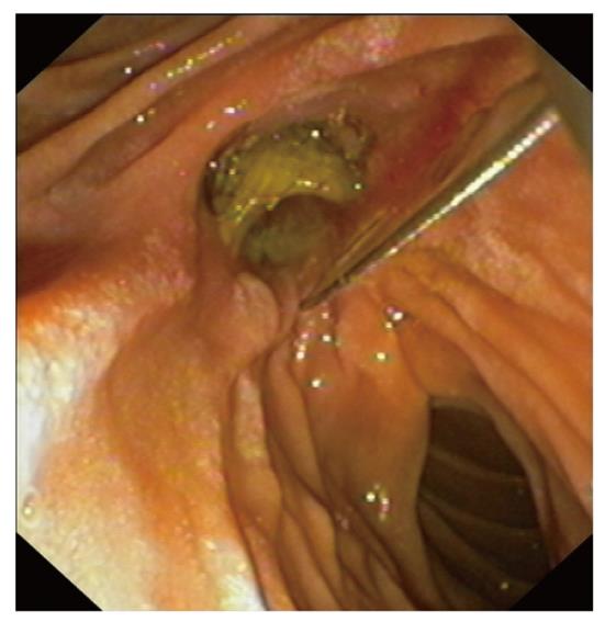
Figure 3 The laceration is evident just below the lower end of the bile duct.
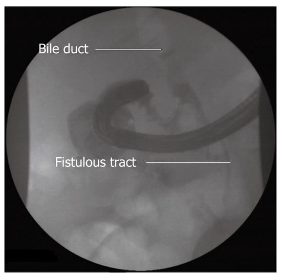
Figure 4 Injection of contrast from the endoscope enables visualization of the fistulous tract.
The bile duct is also delineated with the presence of gas.
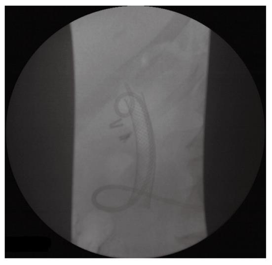
Figure 5 The covered self-expandable metallic biliary stent covers the laceration.
Figure 6 Covered self-expandable metallic biliary stent and nasobiliary catheter in place.
- Citation: Vezakis A, Fragulidis G, Nastos C, Yiallourou A, Polydorou A, Voros D. Closure of a persistent sphincterotomy-related duodenal perforation by placement of a covered self-expandable metallic biliary stent. World J Gastroenterol 2011; 17(40): 4539-4541
- URL: https://www.wjgnet.com/1007-9327/full/v17/i40/4539.htm
- DOI: https://dx.doi.org/10.3748/wjg.v17.i40.4539


