Copyright
©2011 Baishideng Publishing Group Co.
World J Gastroenterol. Oct 14, 2011; 17(38): 4314-4320
Published online Oct 14, 2011. doi: 10.3748/wjg.v17.i38.4314
Published online Oct 14, 2011. doi: 10.3748/wjg.v17.i38.4314
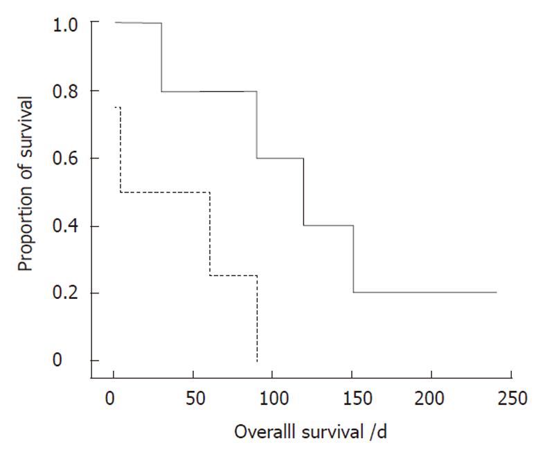
Figure 1 Kaplan-Meier plot of the survival curve for nine patients according to the diagnostic time of gastrointestinal metastasis before surgery (solid line) or after surgery (dashed line).
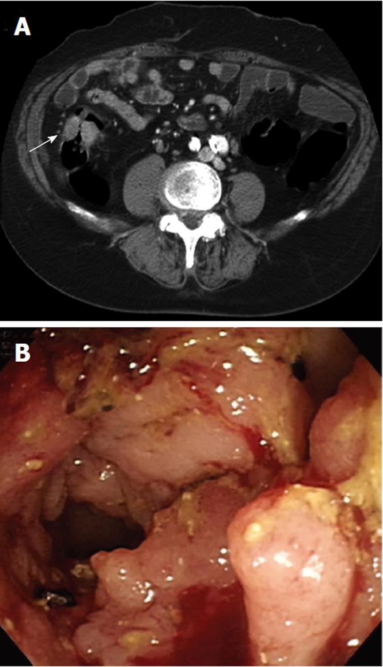
Figure 2 Imaging study of one patient with colonic metastases from lung cancer.
A: An abdominal computed tomography showed a 4-cm ill-defined tumor at the ascending colon (arrow); B: Colonoscopic feature of a circular mass with luminal stenosis and easy-touch bleeding.
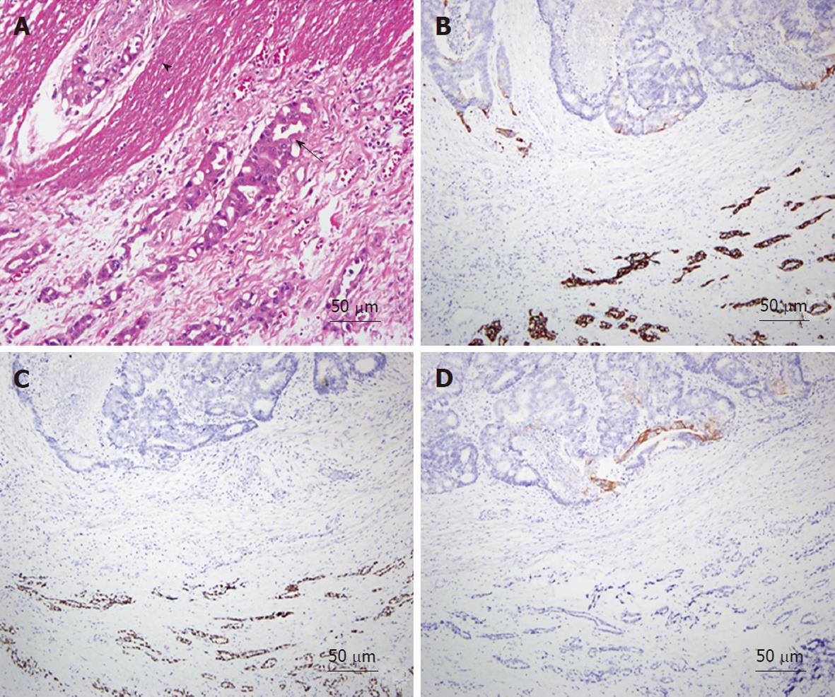
Figure 3 Microscopic findings of metastatic lung adenocarcinoma in the colon.
A: Hematoxylin-eosin staining; × 200. This tumor was characterized by pleomorphic oval nuclei, scant cytoplasm, an irregular glandular pattern (arrow), and invasion of the muscle layer (arrow head) of the colon; B-D: Staining for thyroid transcription factor (TTF)-1 (B), cytokeratin (CK)7 (C), CK20 (D); × 100. In general, CK20 positive means that the tumor originates from the gastrointestinal tract. By immunohistochemistry, the tumor cells located in the lower part of the figure were found to be positive for TTF-1 (B) and CK7 (C), but negative for CK20 (D). The glandular structures in the upper part belong to the colon mucosa.
- Citation: Lee PC, Lo C, Lin MT, Liang JT, Lin BR. Role of surgical intervention in managing gastrointestinal metastases from lung cancer. World J Gastroenterol 2011; 17(38): 4314-4320
- URL: https://www.wjgnet.com/1007-9327/full/v17/i38/4314.htm
- DOI: https://dx.doi.org/10.3748/wjg.v17.i38.4314


