Copyright
©2011 Baishideng Publishing Group Co.
World J Gastroenterol. Oct 14, 2011; 17(38): 4289-4297
Published online Oct 14, 2011. doi: 10.3748/wjg.v17.i38.4289
Published online Oct 14, 2011. doi: 10.3748/wjg.v17.i38.4289
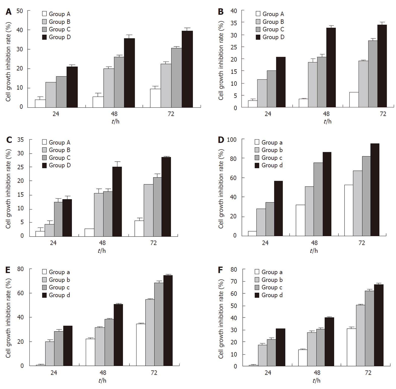
Figure 1 Treatment with rAd-p53 or oxaliplatin alone inhibits the growth of gastric cancer cells in a time- and dose-dependent manner.
SGC-7910 (A), BGC-823 (B) and HGC-27 (C) cells were treated with rAd-p53 followed by the determination of cell growth inhibition rates. Groups A, B, C and D were treated with the indicated rAd-p53 dose (vp/mL) of 5 × 10⁶, 5 × 10⁷, 5 × 10⁸ and 5 × 10⁹, respectively. SGC-7910 (D), BGC-823 (E) and HGC-27 (F) cells were treated with oxaliplatin (OXA), and cell growth inhibition rates were determined. Groups a, b, c and d were treated with the indicated OXA dose (μg/mL) of 3.2, 6.4, 12.8 and 25.6, respectively.
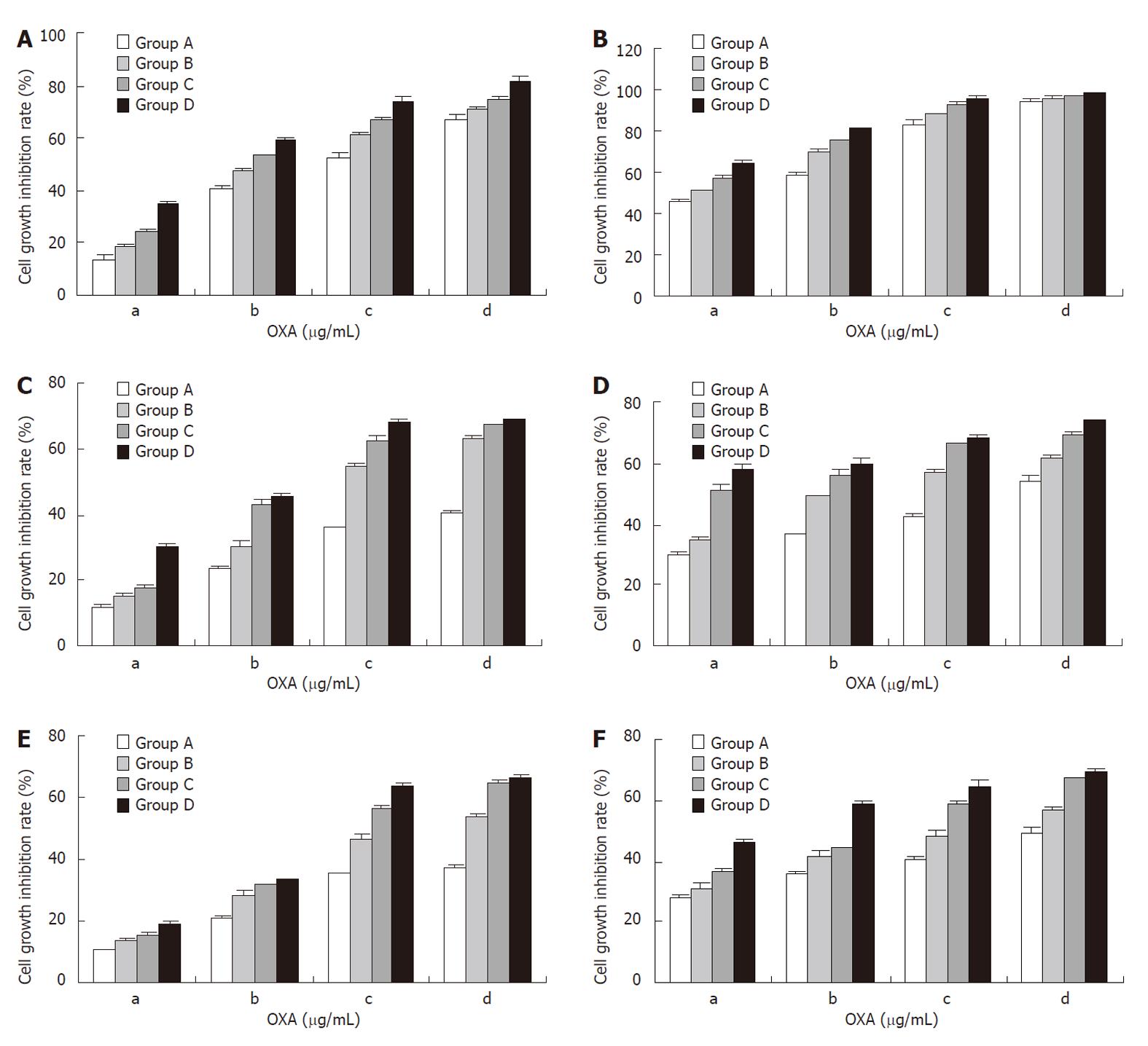
Figure 2 Combination treatment with rAd-p53 and oxaliplatin has synergistic effects on the inhibition of gastric cancer cell growth.
SGC-7910 (A, B), BGC-823 (C, D) and HGC-27 (E, F) cells were treated with rAd-p53 plus oxaliplatin (OXA), and cell growth inhibition rates were determined at 24 h (A, C, E) or 48 h (B, D, F). Groups A, B, C and D were treated with the indicated rAd-p53 dose (vp/mL) of 5 × 10⁶, 5 × 10⁷, 5 × 10⁸ and 5 × 10⁹, respectively. Groups a, b, c and d were treated with the indicated OXA dose (μg/mL) of 3.2, 6.4, 12.8 and 25.6, respectively.
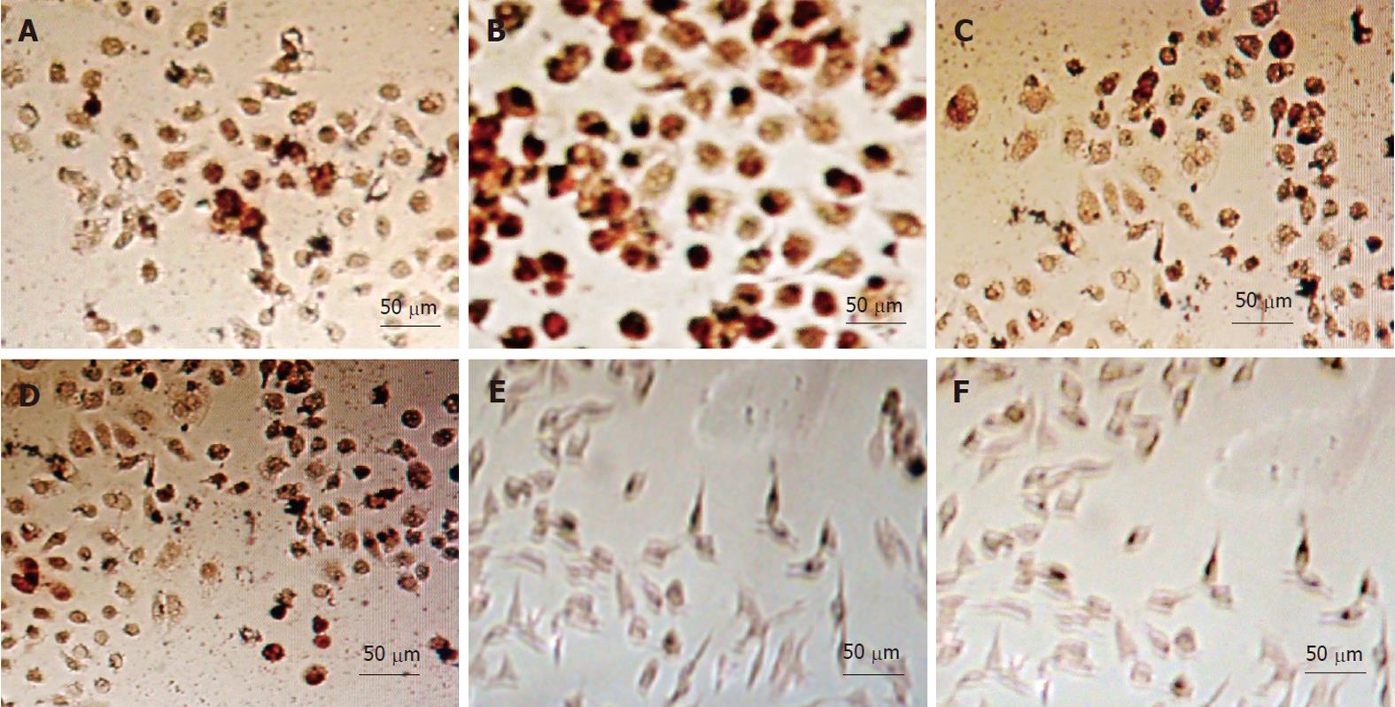
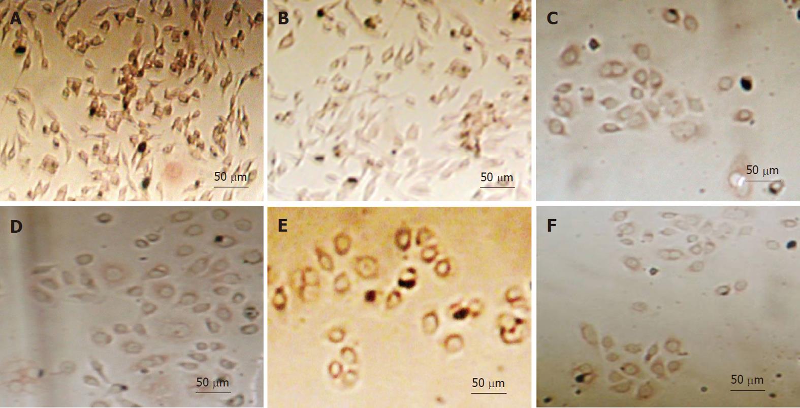
Figure 3 Detection of p53 expression in gastric cancer cells with immunohistochemistry.
A: Untreated SGC-7901 cells; B: SGC-7901 cells treated with 5 × 10⁹ vp/mL rAd-p53 plus 3.2 μg/mL oxaliplatin (OXA); C: Untreated BGC-823 cells; D: BGC-823 cells treated with 5 × 10⁹ vp/mL rAd-p53 plus 3.2 μg/mL OXA; E: Untreated HGC-27 cells untreated; F: HGC-27 cells treated with 5 × 10⁹ vp/mL rAd-p53 plus 3.2 μg/mL OXA.
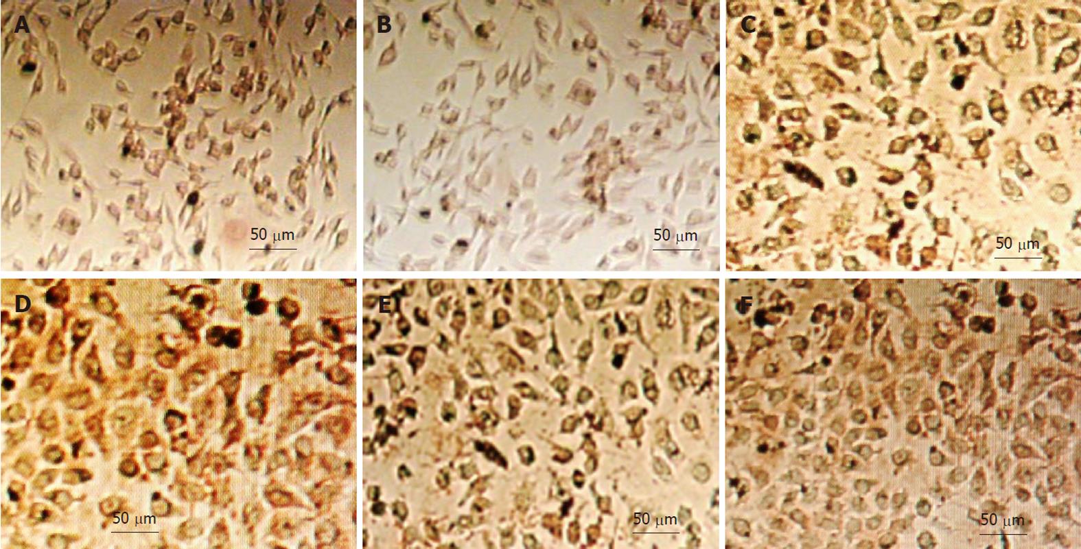
Figure 4 Detection of bax expression in gastric cancer cells with immunohistochemistry.
A: Untreated SGC-7901 cells; B: SGC-7901 cells treated with 5 × 10⁹ vp/mL rAd-p53 plus 3.2 μg/mL oxaliplatin (OXA); C: Untreated BGC-823 cells; D: BGC-823 cells treated with 5 × 10⁹ vp/mL rAd-p53 plus 3.2 μg/mL OXA; E: Untreated HGC-27 cells; F: HGC-27 cells treated with 5 × 10⁹ vp/mL rAd-p53 plus 3.2 μg/mL OXA.
Figure 5 Detection of Bcl-2 expression in gastric cancer cells with immunohistochemistry.
A: Untreated SGC-7901 cells; B: SGC-7901 cells treated with 5 × 10⁹ vp/mL rAd-p53 plus 3.2 μg/mL oxaliplatin (OXA); C: Untreated BGC-823 cells; D: BGC-823 cells treated with 5 × 10⁹ vp/mL rAd-p53 plus 3.2 μg/mL OXA; E: Untreated HGC-27 cells; F: HGC-27 cells treated with 5 × 10⁹ vp/mL rAd-p53 plus 3.2 μg/mL OXA.
- Citation: Chen GX, Zheng LH, Liu SY, He XH. rAd-p53 enhances the sensitivity of human gastric cancer cells to chemotherapy. World J Gastroenterol 2011; 17(38): 4289-4297
- URL: https://www.wjgnet.com/1007-9327/full/v17/i38/4289.htm
- DOI: https://dx.doi.org/10.3748/wjg.v17.i38.4289


