Copyright
©2011 Baishideng Publishing Group Co.
World J Gastroenterol. Sep 21, 2011; 17(35): 4013-4016
Published online Sep 21, 2011. doi: 10.3748/wjg.v17.i35.4013
Published online Sep 21, 2011. doi: 10.3748/wjg.v17.i35.4013
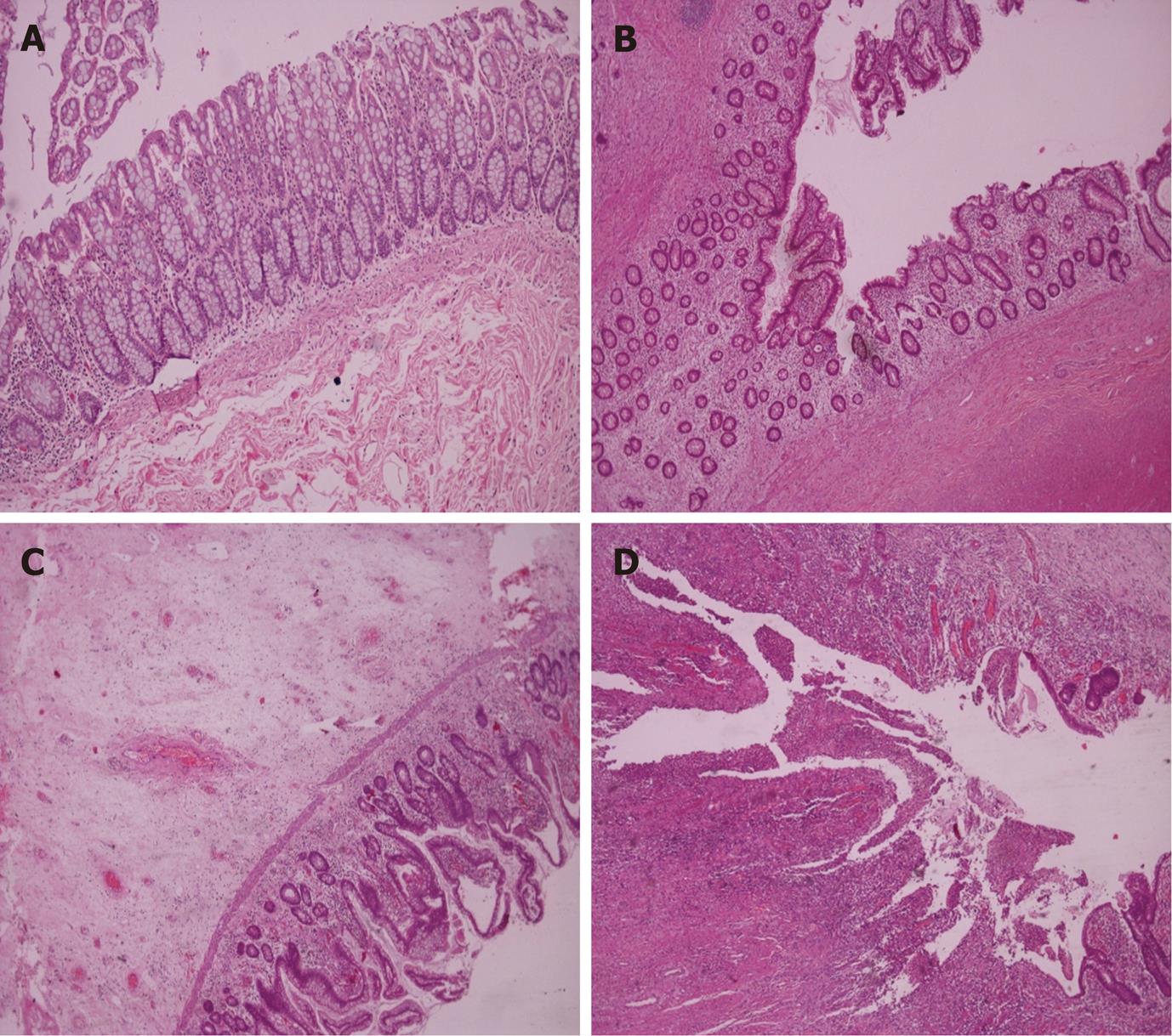
Figure 1 Representative histologic findings of normal (A) and radiation enteritis associated (B-D) intestinal mucosa.
A: Normal; B: Mild lesions with edema and increased number of fibroblasts; C: Moderate lesions with sub-mucosal edema and disturbed mucosal architecture; D: Severe lesions with mucosal ulceration. Tissue sections were stained with standard hematoxylin-eosin (40 x magnification).
- Citation: Perrakis N, Athanassiou E, Vamvakopoulou D, Kyriazi M, Kappos H, Vamvakopoulos NC, Nomikos I. Practical approaches to effective management of intestinal radiation injury: Benefit of resectional surgery. World J Gastroenterol 2011; 17(35): 4013-4016
- URL: https://www.wjgnet.com/1007-9327/full/v17/i35/4013.htm
- DOI: https://dx.doi.org/10.3748/wjg.v17.i35.4013


