Copyright
©2011 Baishideng Publishing Group Co.
World J Gastroenterol. Sep 21, 2011; 17(35): 3962-3970
Published online Sep 21, 2011. doi: 10.3748/wjg.v17.i35.3962
Published online Sep 21, 2011. doi: 10.3748/wjg.v17.i35.3962
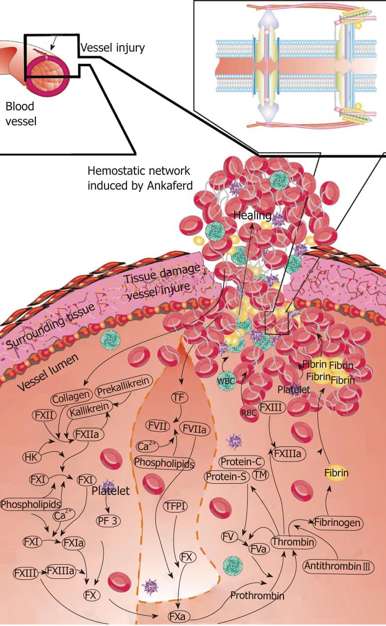
Figure 1 The basic mechanism of action for ankaferd blood stopper is the formation of an encapsulated protein network that provides focal points for erythrocyte aggregation.
Ankaferd blood stopper (ABS)-induced formation of the unique protein network within the vital erythroid aggregation covers the entire physiological haemostatic process. Red blood cell (RBC) elements (such as spectrin and ankrin surface receptors, and internal ferrochelatase enzyme), related transcription factors (such as GATA-1) and RBC-related proteins (such as urotensin II) are the main targets of ABS. Those proteins and the required adenosine triphosphate bioenergy are included in the protein library of Ankaferd[1].
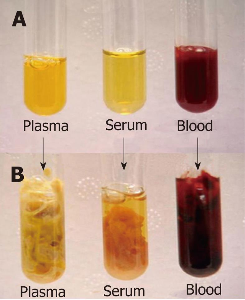
Figure 2 Ankaferd blood stopper-induced protein network formation within less than 1 s.
Plasma under the light microscopy before (A) and just after (B) Ankaferd application[1].
Figure 3 Ancaferd application during endoscopic sphincterotomy.
A: Early bleeding during the endoscopic sphincterotomy; B: Ankaferd blood stopper (ABS) topically applied to the bleeding area; C: Hemorrhage was immediately controlled just after topical ABS administration; D: The bleeding site was covered by the hemostatic network related with the ABS application and hemorrhage was stopped.
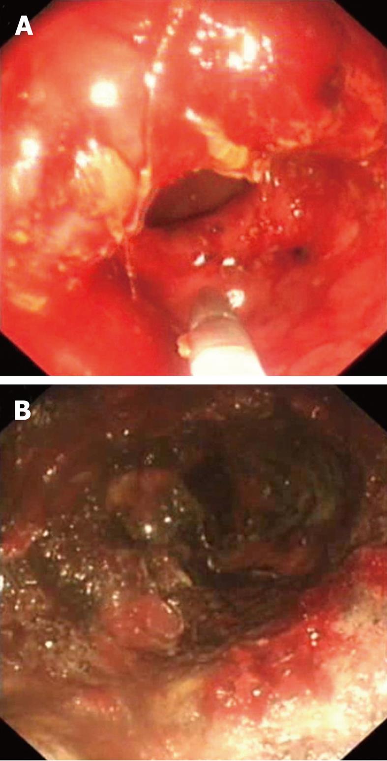
Figure 4 Endocopic images of the distal rectum in a patient with radiation proctitis.
A: Before Ankaferd blood stopper (ABS) application with fresh bleeding; B: After ABS application with grayish-yellow coagulum formation covering the diseased area.
- Citation: Beyazit Y, Kekilli M, Haznedaroglu IC, Kayacetin E, Basaranoglu M. Ankaferd hemostat in the management of gastrointestinal hemorrhages. World J Gastroenterol 2011; 17(35): 3962-3970
- URL: https://www.wjgnet.com/1007-9327/full/v17/i35/3962.htm
- DOI: https://dx.doi.org/10.3748/wjg.v17.i35.3962


