Copyright
©2011 Baishideng Publishing Group Co.
World J Gastroenterol. Sep 14, 2011; 17(34): 3953-3956
Published online Sep 14, 2011. doi: 10.3748/wjg.v17.i34.3953
Published online Sep 14, 2011. doi: 10.3748/wjg.v17.i34.3953
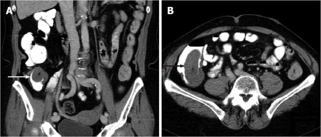
Figure 1 Coronary (A) and transversal coupe (B) of abdominal computed tomography in a patient with neurofibromatosis type 1 with a cecal mass (arrows).
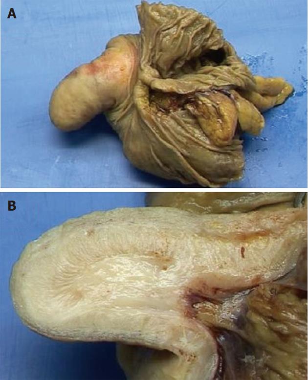
Figure 2 Opened resection specimen (A) and cut surface of the polyp (B).
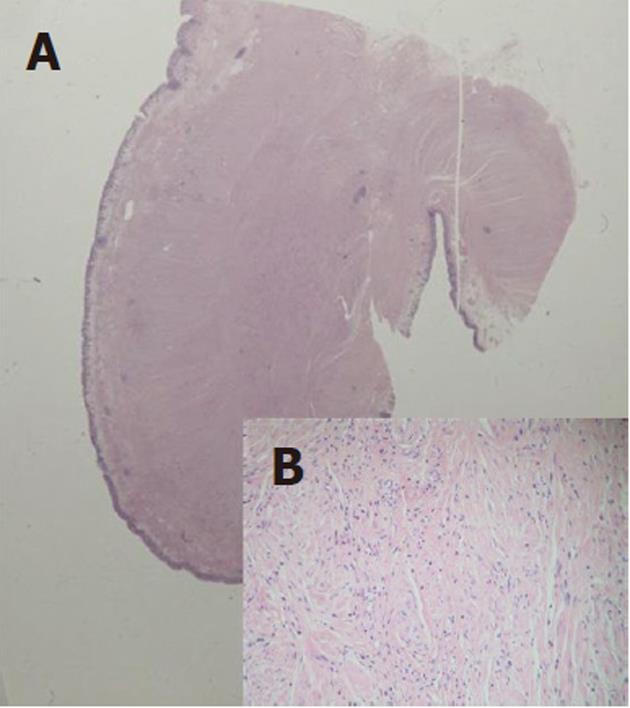
Figure 3 Overview of the polyp (A) and detail of the submucosal tumor (B), HE staining; original magnification, × 100.
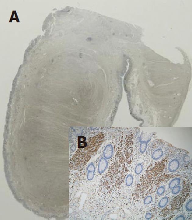
Figure 4 Overview of the polyp (A) and detail of the submucosal tumor (B), S100 staining; original magnification, × 100.
- Citation: Donk W, Poyck P, Westenend P, Lesterhuis W, Hesp F. Recurrent abdominal complaints caused by a cecal neurofibroma: A case report. World J Gastroenterol 2011; 17(34): 3953-3956
- URL: https://www.wjgnet.com/1007-9327/full/v17/i34/3953.htm
- DOI: https://dx.doi.org/10.3748/wjg.v17.i34.3953


