Copyright
©2011 Baishideng Publishing Group Co.
World J Gastroenterol. Sep 14, 2011; 17(34): 3933-3940
Published online Sep 14, 2011. doi: 10.3748/wjg.v17.i34.3933
Published online Sep 14, 2011. doi: 10.3748/wjg.v17.i34.3933
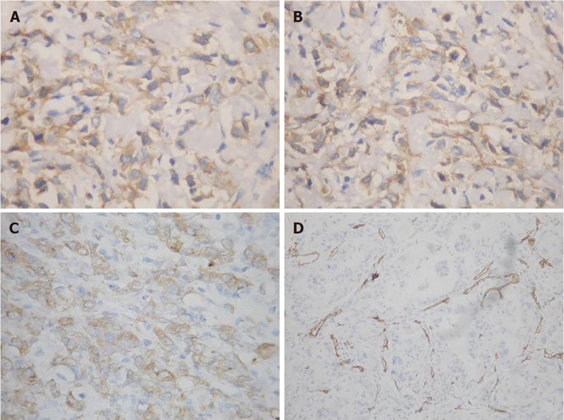
Figure 1 Erythropoietin, erythropoietin receptor, vascular endothelial growth factor and CD34 expression in biopsies from gastric adenocarcinoma patients.
A, B: Erythropoietin and erythropoietin receptor positive immunostaining signals were localized in the cytoplasm of tumor cells with a scattered distribution pattern in most cases; C: Vascular endothelial growth factor immunoreactivity was detected in the cytoplasm of gastric adenocarcinoma. Original magnification: × 200; D: Microvessels in tumor were highlighted by staining endothelial cells for anti-CD34. Any brown-staining endothelial cell or cell cluster that was clearly separated from adjacent microvessels was considered as a single, countable microvessel. Original magnification: × 100.
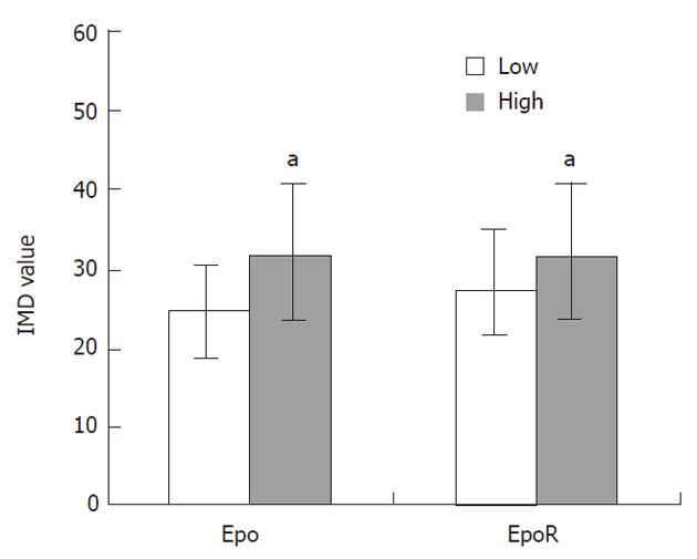
Figure 2 Correlation between intratumoral microvessel density and protein expression in gastric adenocarcinoma.
The mean intratumoral microvessel density in the high-expressed erythropoietin (Epo) and erythropoietin receptor (EpoR) cases was significantly increased compared to that of low-expressed cases (aP < 0.05). IMD: Intratumoral microvessel density.
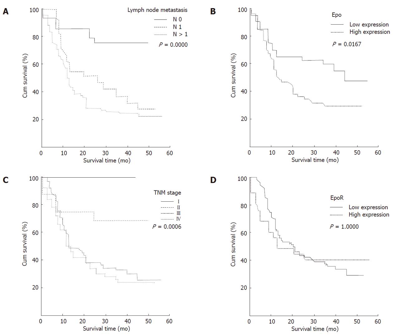
Figure 3 Kaplan-Meier survival analyses of gastric adenocarcinoma patients.
A: Kaplan-Meier curve showing that patients with lymph node metastasis (N ≥ 1) have a lower survival rate than those without lymph node metastasis (N0); B: A significant difference in survival rate was found between gastric adenocarcinoma (GAC) patients with high and low erythropoietin expression in tumor; C: A significant difference in survival rate was found between different clinical stages; D: No significant difference in survival rate was found between GAC patients with high and low erythropoietin receptor expression in tumor.
- Citation: Wang L, Li HG, Xia ZS, Wen JM, Lv J. Prognostic significance of erythropoietin and erythropoietin receptor in gastric adenocarcinoma. World J Gastroenterol 2011; 17(34): 3933-3940
- URL: https://www.wjgnet.com/1007-9327/full/v17/i34/3933.htm
- DOI: https://dx.doi.org/10.3748/wjg.v17.i34.3933


