Copyright
©2011 Baishideng Publishing Group Co.
World J Gastroenterol. Sep 7, 2011; 17(33): 3802-3809
Published online Sep 7, 2011. doi: 10.3748/wjg.v17.i33.3802
Published online Sep 7, 2011. doi: 10.3748/wjg.v17.i33.3802
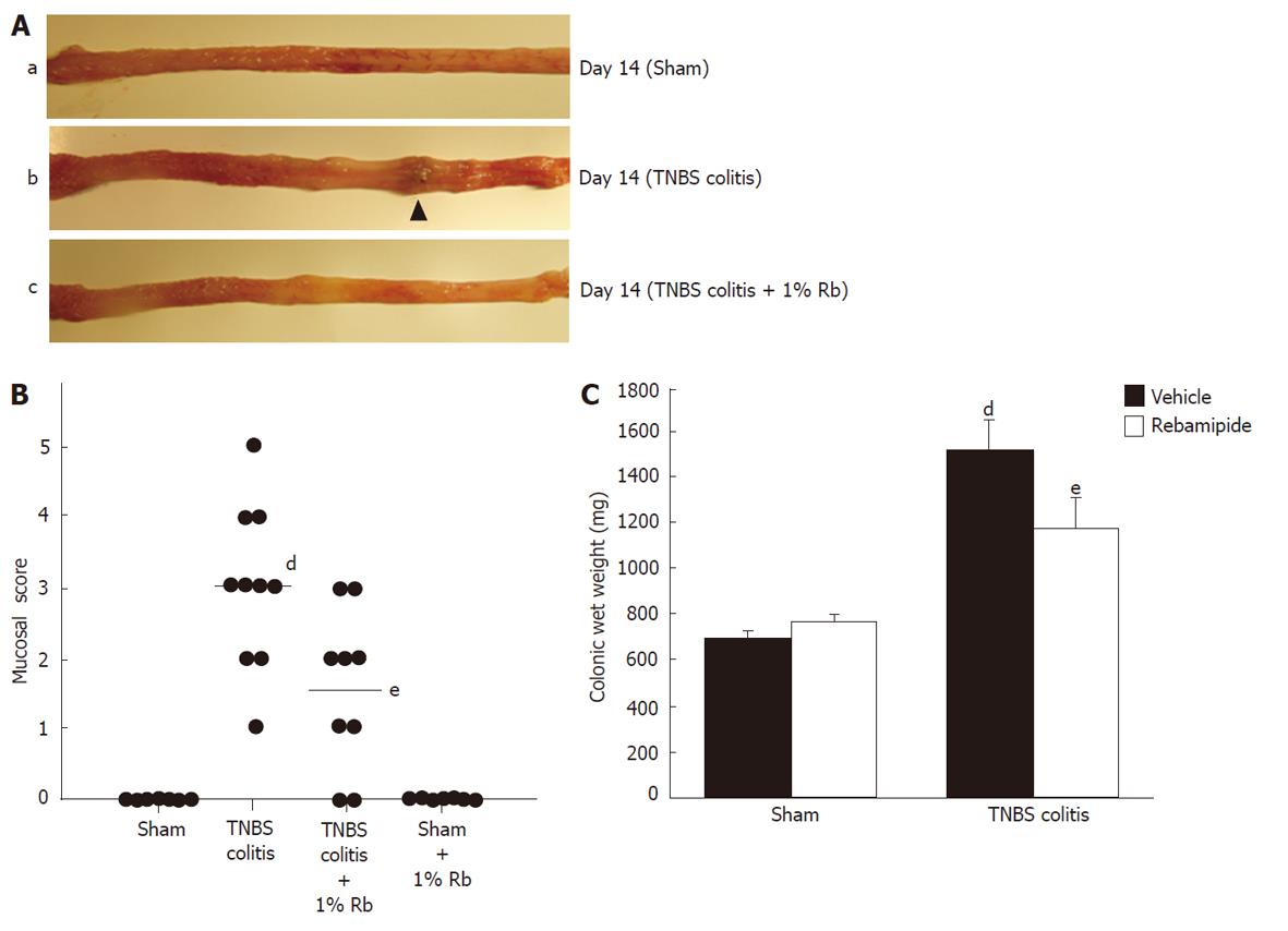
Figure 1 Effects of 1% rebamipide on macroscopic findings, mucosal damage score, and wet colon weight on day 14 after trinitrobenzene sulfonic acid-induced injury.
A: Severe colitis was induced with hyperemia, edema, thickening, ulceration, and necrosis in trinitrobenzene sulfonic acid (TNBS)-colitis rats (b) compared to sham-operated rats (a). These changes were reduced in rats treated with 1% rebamipide (TNBS-colitis rats treated with 1% rebamipide) (c). B: A 1% rebamipide enema was administrated twice daily starting on day 7 after induction of colitis, until day 14. Rats were sacrificed on day 14, and the mucosal damage score was evaluated. Data are expressed as a scatter plot. dP < 0.01 vs sham-treated rats. eP < 0.05 vs TNBS-induced colitis rats receiving the vehicle. C: Rats were sacrificed on day 14 and the distal colon was removed, after which, the wet colon weight was immediately measured. Data represent the mean ± SE of seven rats. dP < 0.01 vs sham-treated rats receiving the vehicle. eP < 0.01 vs TNBS-induced colitis rats receiving the vehicle.
Figure 2 Effects of 1% rebamipide on histological findings in the colon on day 14 after trinitrobenzene sulfonic acid-induced injury.
A: Histological appearance of colonic tissue in sham-operated rats (a), trinitrobenzene sulfonic acid (TNBS)-colitis rats (b), and TNBS-colitis rats treated with 1% rebamipide (c). Histological examination revealed that TNBS administration induced marked thickening of the colonic wall, which was associated with transmural infiltration of inflammatory cells. In contrast, both mural wall thickening and infiltration of inflammatory cells were inhibited in rats treated with 1% rebamipide. Hematoxylin and eosin staining (× 40). B: Histological score was evaluated. Data represent the mean ± SE of six rats. dP < 0.01 vs TNBS-colitis rats receiving the vehicle.
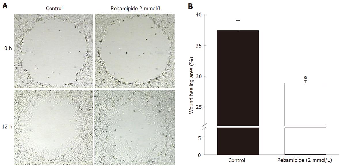
Figure 3 Restitution of rat intestinal epithelial cells around an artificially created wound in control and rebamipide-treated groups.
A: Restitution of rat intestinal epithelial (RIE) cells was evaluated using a wound assay. The denuded area of RIE cells recovered in a time-dependent manner after wound induction. Restitution of the denuded area was promoted by rebamipide at 12 h after wound induction. B: To investigate the effects of rebamipide on RIE cell migration, cells were co-incubated with 2 mmol/L rebamipide after wound induction. Wound repair in 2 mmol/L rebamipide-treated cells occurred significantly earlier than it did in the controls. Datas represent the mean ± SE of four experiments. aP < 0.05 vs controls.
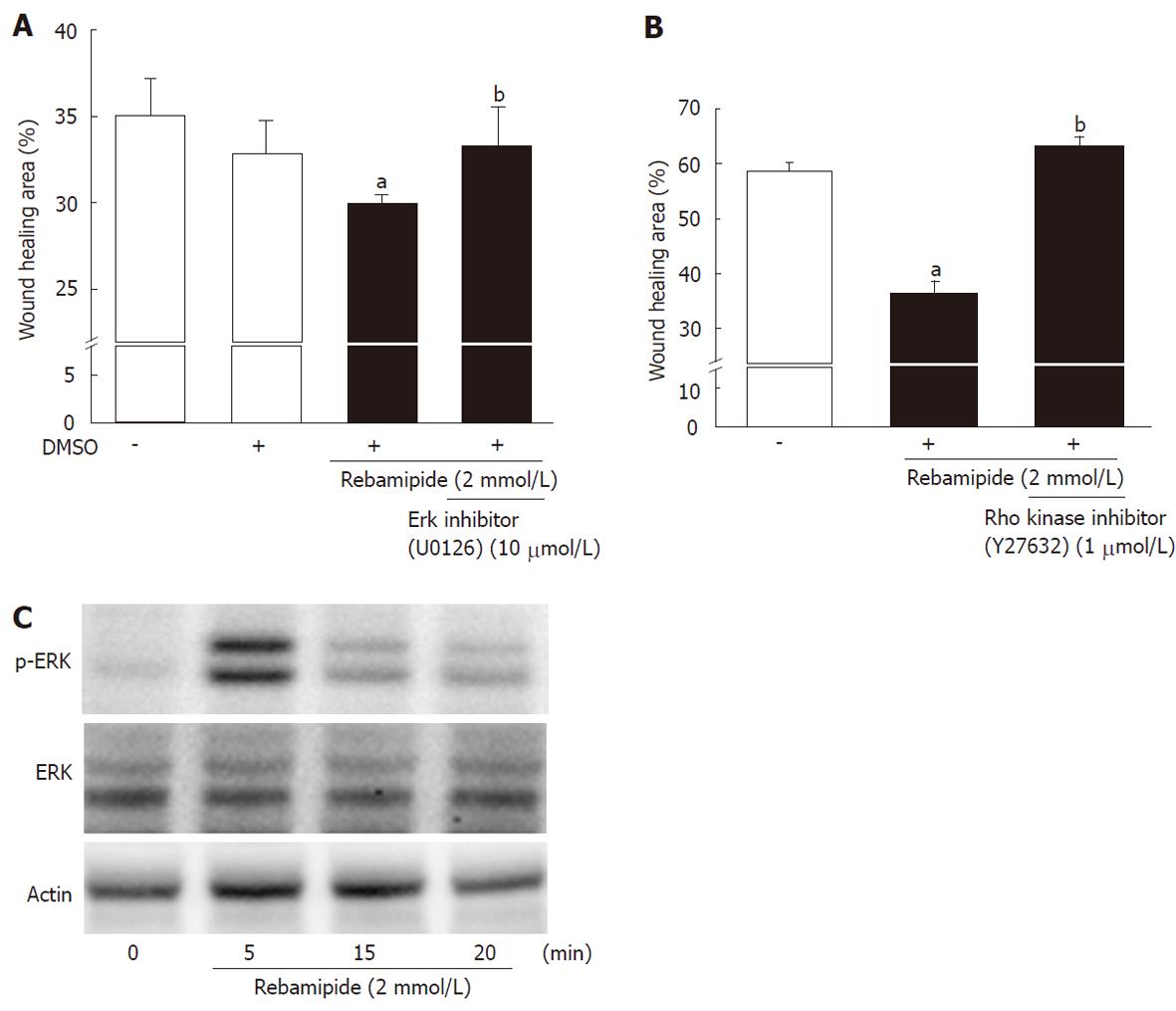
Figure 4 Involvement of extracellular signal-regulated kinase and Rho in rebamipide-treated rat intestinal epithelial cells.
A: Rat intestinal epithelial (RIE) cells were treated with rebamipide (2 mmol/L) with or without an extracellular signal-regulated kinase inhibitor (U01263, 10 μmol/L) after wound induction. The wound healing area (12 h later) was then monitored. Data represent the mean ± SE of four experiments. aP < 0.05 vs controls. bP < 0.05 vs the 2 mmol/Lrebamipide-treated group. B: Expression of p44/42 and phospho-p44/42 in RIE cells incubated with rebamipide (2 mmol/L) was measured using western blot analysis. Actin antibody was used as an internal control. Representative data from three observations is shown. C: RIE cells were treated with rebamipide (2 mmol/L) with or without Rho kinase inhibitor (Y27632, 1 μmol/L) after wound induction. The wound healing area (6 h later) was then monitored. Data represent the mean ± SE of four experiments. aP < 0.05 vs controls. bP < 0.05 vs the 2 mmol/L rebamipide-treated group. DMSO: Dimethyl sulfoxide.
- Citation: Takagi T, Naito Y, Uchiyama K, Okuda T, Mizushima K, Suzuki T, Handa O, Ishikawa T, Yagi N, Kokura S, Ichikawa H, Yoshikawa T. Rebamipide promotes healing of colonic ulceration through enhanced epithelial restitution. World J Gastroenterol 2011; 17(33): 3802-3809
- URL: https://www.wjgnet.com/1007-9327/full/v17/i33/3802.htm
- DOI: https://dx.doi.org/10.3748/wjg.v17.i33.3802


