Copyright
©2011 Baishideng Publishing Group Co.
World J Gastroenterol. Aug 28, 2011; 17(32): 3745-3751
Published online Aug 28, 2011. doi: 10.3748/wjg.v17.i32.3745
Published online Aug 28, 2011. doi: 10.3748/wjg.v17.i32.3745
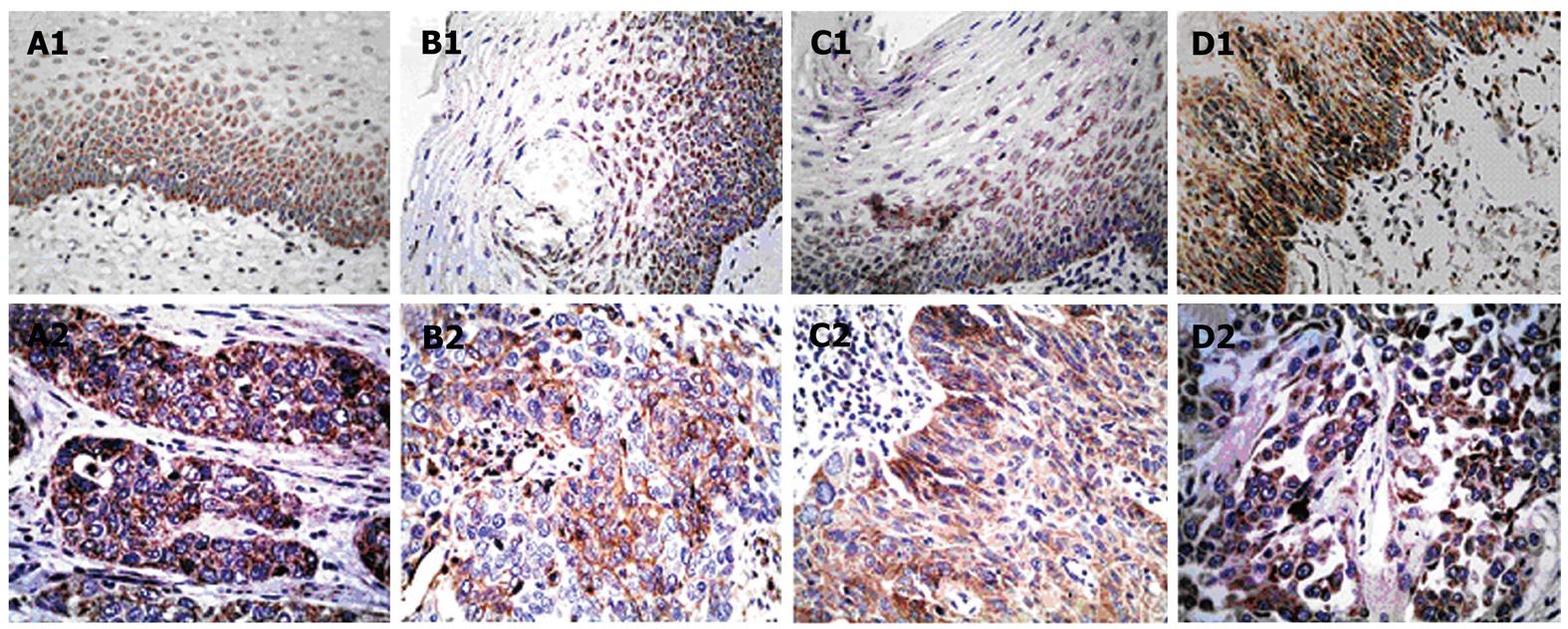
Figure 1 Immunohistochemistry staining of esophageal lesions with Toll-like receptor-specific mAbs.
A1 to D1 show the expression of Toll-like receptor (TLR) 3, TLR4, TLR7 and TLR9 in normal esophageal epithelium, respectively. A2 and C2 show positive staining for TLR3 and TLR7 in esophageal squamous cell carcinoma cells. B2 shows positive TLR4 staining in tumor cells and mononuclear inflammatory cells, and D2 shows positive TLR9 staining in tumor cells and fibroblast-like cells (Original magnification, × 400).
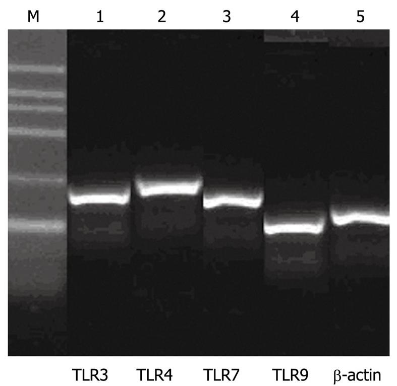
Figure 2 mRNA expression patterns of Toll-like receptor 3, Toll-like receptor 4, Toll-like receptor 7 and Toll-like receptor 9.
M: 100-600 bp marker ladder. Lanes 1 to 4 show the expression of Toll-like receptors, and lane 5 shows the expression of β-actin.
- Citation: Sheyhidin I, Nabi G, Hasim A, Zhang RP, Ainiwaer J, Ma H, Wang H. Overexpression of TLR3, TLR4, TLR7 and TLR9 in esophageal squamous cell carcinoma. World J Gastroenterol 2011; 17(32): 3745-3751
- URL: https://www.wjgnet.com/1007-9327/full/v17/i32/3745.htm
- DOI: https://dx.doi.org/10.3748/wjg.v17.i32.3745


