Copyright
©2011 Baishideng Publishing Group Co.
World J Gastroenterol. Aug 14, 2011; 17(30): 3531-3537
Published online Aug 14, 2011. doi: 10.3748/wjg.v17.i30.3531
Published online Aug 14, 2011. doi: 10.3748/wjg.v17.i30.3531
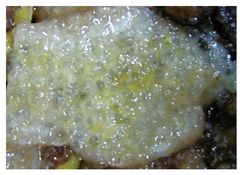
Figure 1 Upon sectioning, the peritoneal lesions were full of a jelly-like mucous substance.
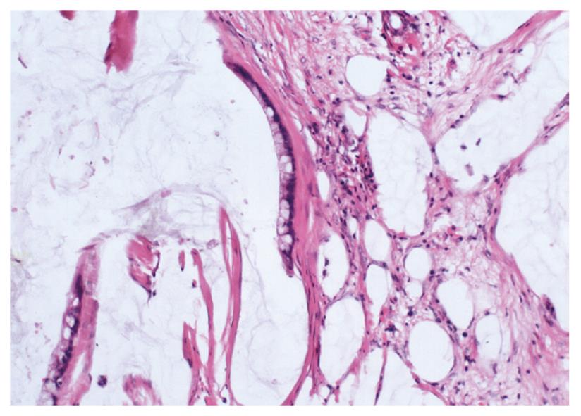
Figure 2 Microscopically, some mucous glandular structures were floating in a large number of mucous lakes (HE stain, × 200).
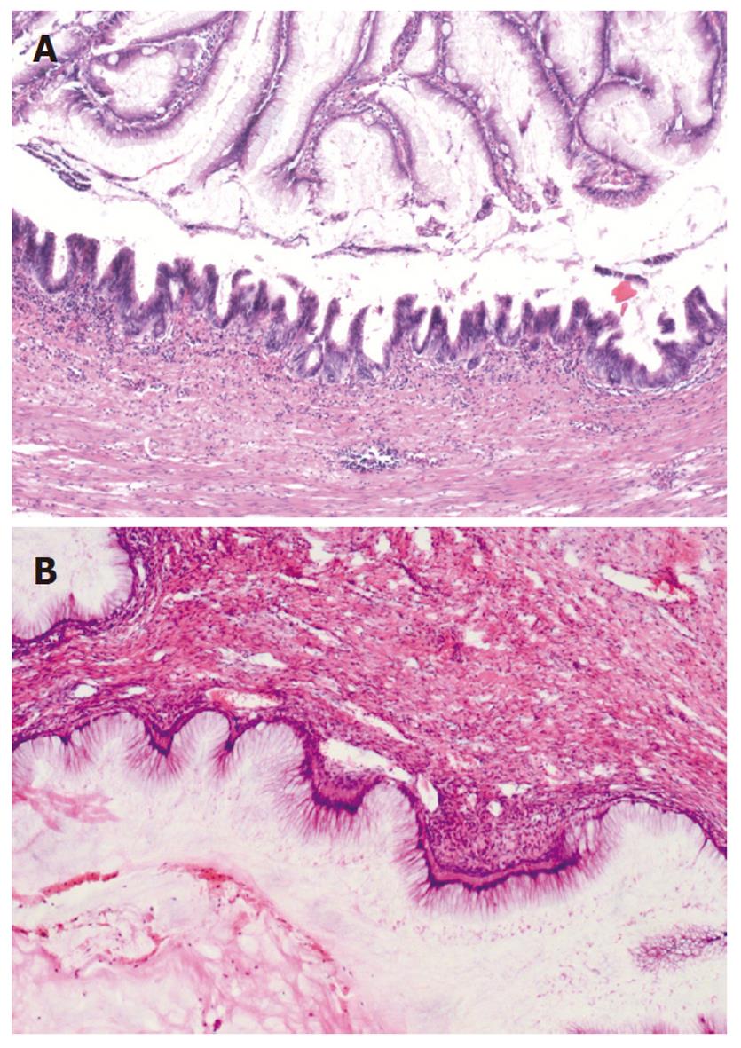
Figure 3 Mucinous tumors were observed in the appendix (A) (HE stain, ×100) and ovary (B) (HE stain, × 100).
Figure 4 Mucin-2 was positive in peritoneal (A) (× 200), appendiceal (B) (× 100) and ovarian (C) (× 100) lesions.
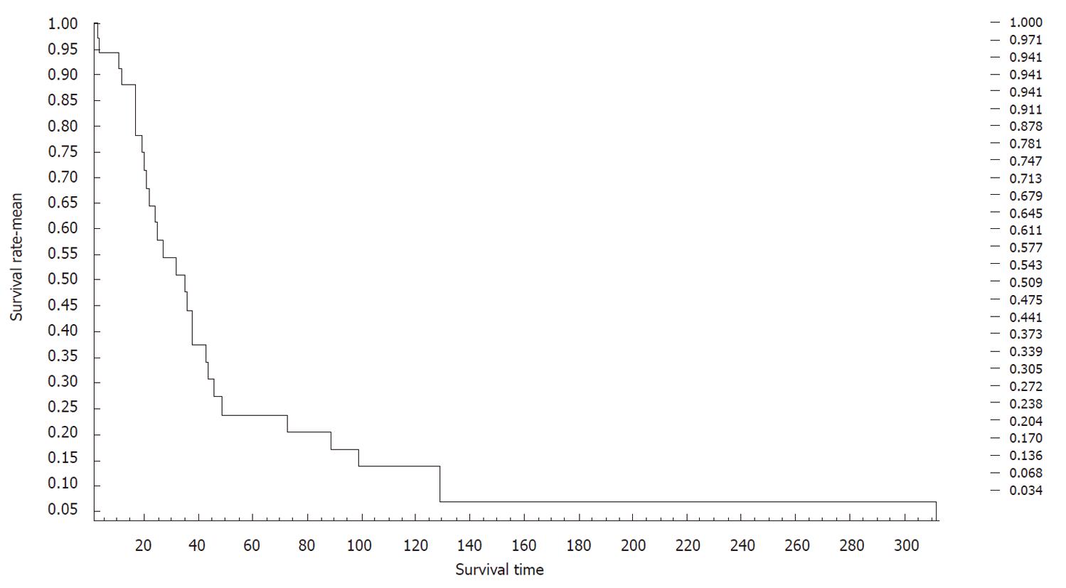
Figure 5 Survival of 35 women with pseudomyxoma peritonei.
- Citation: Guo AT, Song X, Wei LX, Zhao P. Histological origin of pseudomyxoma peritonei in Chinese women: Clinicopathology and immunohistochemistry. World J Gastroenterol 2011; 17(30): 3531-3537
- URL: https://www.wjgnet.com/1007-9327/full/v17/i30/3531.htm
- DOI: https://dx.doi.org/10.3748/wjg.v17.i30.3531


