Copyright
©2011 Baishideng Publishing Group Co.
World J Gastroenterol. Jan 21, 2011; 17(3): 379-384
Published online Jan 21, 2011. doi: 10.3748/wjg.v17.i3.379
Published online Jan 21, 2011. doi: 10.3748/wjg.v17.i3.379
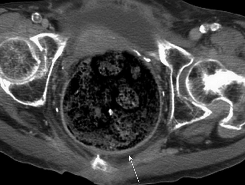
Figure 1 A 70-year-old woman (patient 6) with necrotic stercoral colitis.
The computed tomography scan revealed stool impaction and distension of the recto-sigmoid colon with asymmetrical wall thickening at the posterior aspect (arrow).
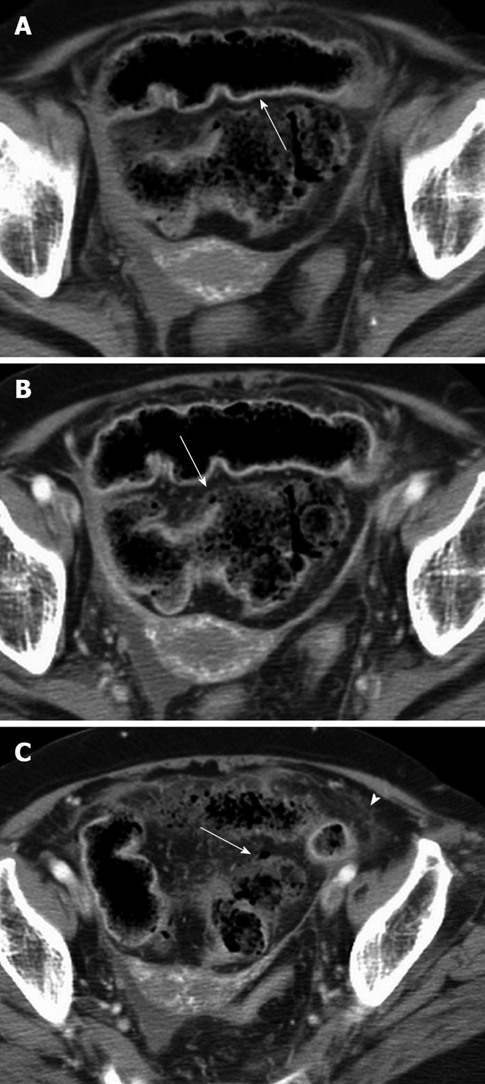
Figure 2 An 80-year-old woman (patient 5) with perforation of the necrotic stercoral colitis at the sigmoid colon.
A: An unenhanced computed tomography (CT) scan reveals dense mucosa (arrow) conforming to the colon wall; B: An enhanced abdominal CT scan reveals discontinuation of the colonic mucosa (arrow) suggesting perfusion defect; C: A small air bubble abutting the damaged colon (arrow) and increased pericolonic infiltration (arrowhead) can be seen.
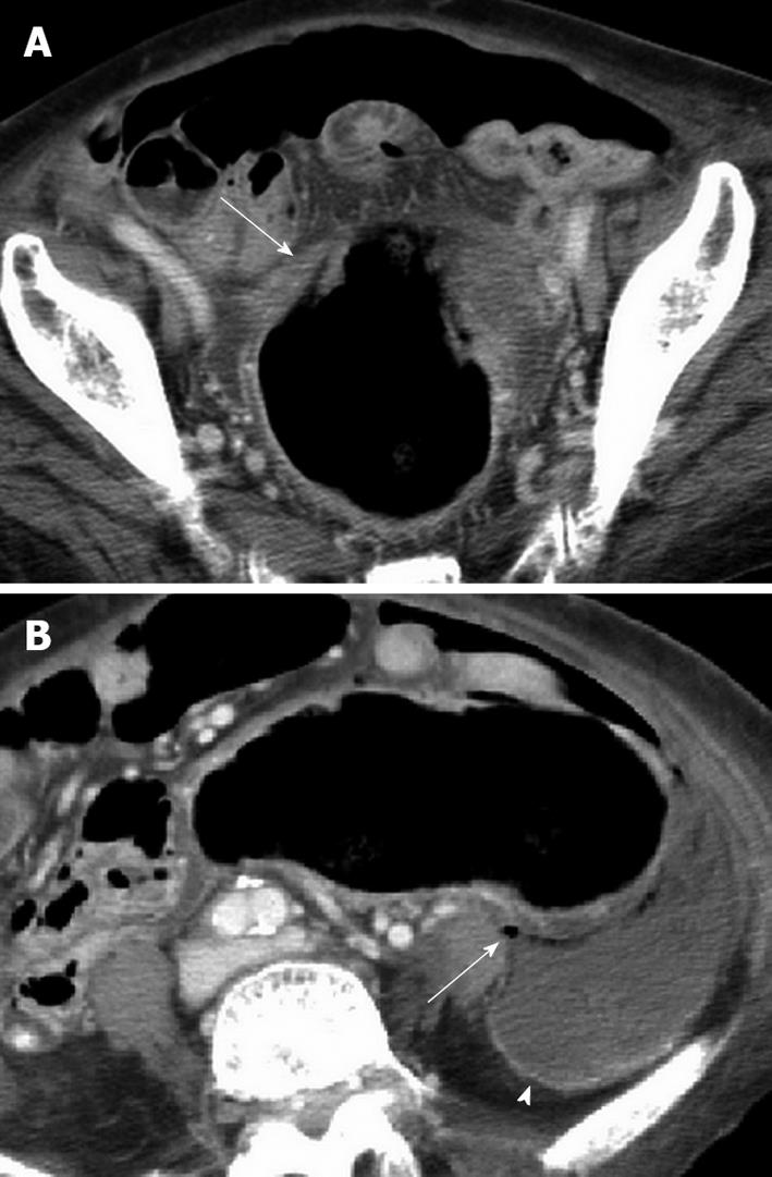
Figure 3 An 88-year-old women (patient 7) with perforation of the necrotic stercoral colitis at the sigmoid colon.
A: An enhanced abdominal computed tomography scan reveals mucosal flap (arrow) slough into the lumen of the colon indicating mucosal sloughing; B: Air pockets (arrow) abutting the colonic wall and pericolonic loculated fluid indicative of abscess formation (arrowhead).
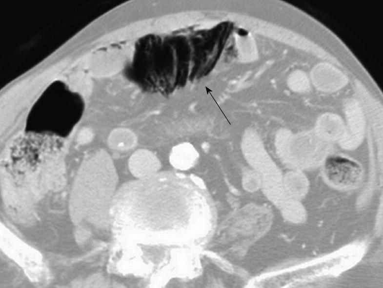
Figure 4 An 87-year-old man (patient 4) with perforated stercoral colitis at the proximal end of co-existing rectosigmoid colon cancer.
An enhanced abdominal computed tomography scan at lung-window setting reveals air confined inside the mesocolon indicating pneumo-mesocolon (arrow).
- Citation: Wu CH, Wang LJ, Wong YC, Huang CC, Chen CC, Wang CJ, Fang JF, Hsueh C. Necrotic stercoral colitis: Importance of computed tomography findings. World J Gastroenterol 2011; 17(3): 379-384
- URL: https://www.wjgnet.com/1007-9327/full/v17/i3/379.htm
- DOI: https://dx.doi.org/10.3748/wjg.v17.i3.379


