Copyright
©2011 Baishideng Publishing Group Co.
World J Gastroenterol. Jun 28, 2011; 17(24): 2972-2975
Published online Jun 28, 2011. doi: 10.3748/wjg.v17.i24.2972
Published online Jun 28, 2011. doi: 10.3748/wjg.v17.i24.2972
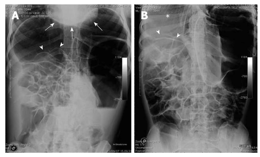
Figure 1 Upright (A) and supine (B) films in a female patient complaining of long-lasting constipation, with recurrent episodes of abdominal pain and distension.
A: An isolated air-fluid level is depicted in the left flank, suggesting mechanical obstruction at the level of the descending colon. Subphrenic radiolucency is evident on both sides, together with the outlining of the intestinal wall of some small bowel loops in the right flank (arrowheads) and that of the inferior cardiac border (arrows); B: Outline of the intestinal wall of small bowel loops appears to be depicted in the right flank (arrowheads), configuring the so-called bas-relief or Rigler’s sign. In addition, hyperlucency is depicted in the right subphrenic space in place of the normal hepatic shadow, configuring the bright or hyperlucent liver sign (*).
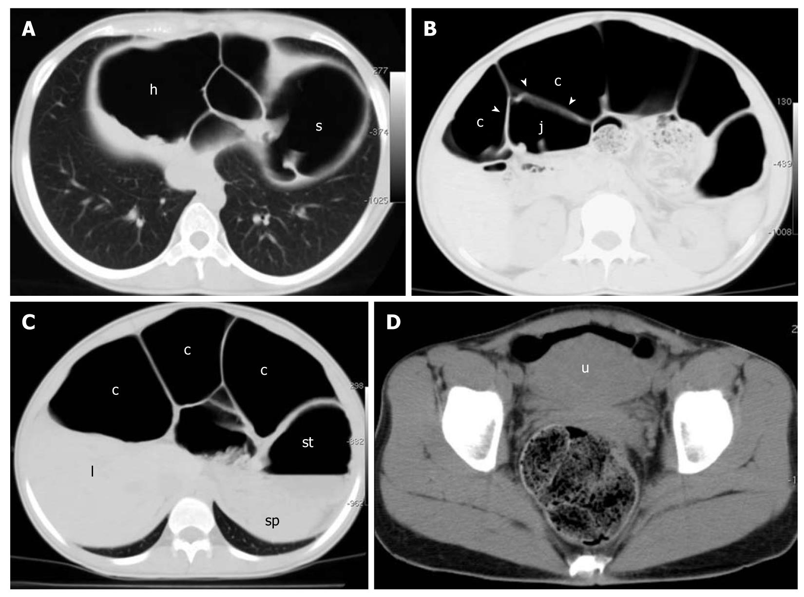
Figure 2 Unenhanced four-row MDCT.
Images are displayed with lung (A-C) and soft tissue (D) windows. A: Hepatic (h) and splenic (s) flexures, air-filled and abnormally dilated, are displaced under the diaphragm, accounting for the subphrenic radiolucency depicted in the upright film; B: Intraluminal air in the large bowel (c) appears to outline the intestinal wall (arrowheads) of a jejunal loop (j) that is also mildly dilated and air-filled, accounting for the Rigler’s sign depicted on the upright and supine films; C: Abnormally dilated colonic segment situated between the liver (l) and anterior abdominal wall, simulating the hyperlucent (bright) liver sign depicted in the supine film; st: Stomach; sp: Spleen; D: Rectum is normally filled with feces. u: Uterus.
- Citation: Camera L, Calabrese M, Sarnelli G, Longobardi M, Rocco A, Cuomo R, Salvatore M. Pseudopneumoperitoneum in chronic intestinal pseudo-obstruction: A case report. World J Gastroenterol 2011; 17(24): 2972-2975
- URL: https://www.wjgnet.com/1007-9327/full/v17/i24/2972.htm
- DOI: https://dx.doi.org/10.3748/wjg.v17.i24.2972


