Copyright
©2011 Baishideng Publishing Group Co.
World J Gastroenterol. Jun 28, 2011; 17(24): 2924-2932
Published online Jun 28, 2011. doi: 10.3748/wjg.v17.i24.2924
Published online Jun 28, 2011. doi: 10.3748/wjg.v17.i24.2924
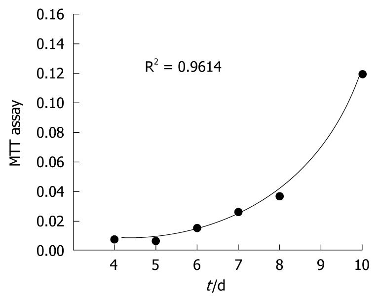
Figure 1 MTT assay.
Twenty-five weeks orally administrated TAA rat liver tissues cells have been harvested and successfully cultured. The cells followed a typical growth curve; lag, logarithmic, and stationary phases during culturing were estimated. Under series surveillance, a cell population doubling time of 32 h was determined (6 points at day 4, 5, 6, 7, 8, and 10).
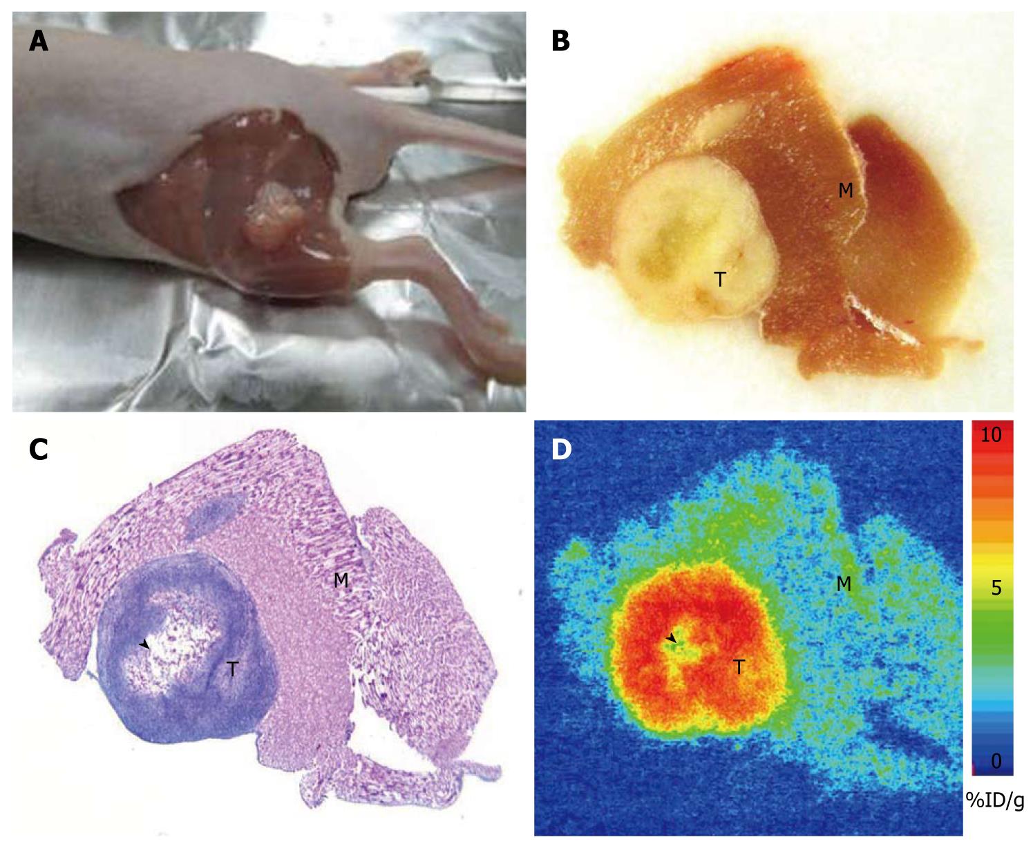
Figure 2 The tumors formed at the cell transplantation site of the CCA xenograft with central necrosis in all large tumors suggesting the fast growth of this malignant tumor at the thigh of the nude mice had a latency period of 4-6 wk and, as represented by (A) gross picture, (B) the photomicrographs of the necropsy, (C) histology, and (D) autoradiography (D).
Of note, all tumor cells excluding necrosis tissue possessed high 2-Deoxy-2-(18F)fluoro-D-glucose uptake.
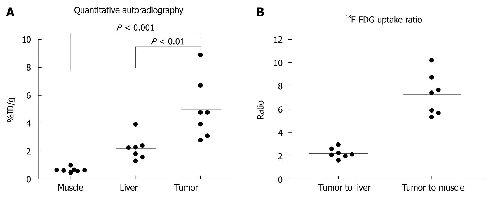
Figure 3 The mean quantitative uptake values of fluorodeoxyglucose in the muscle, liver, and tumor represented by injected dose per gram of tissue (%ID/g) were (A) 0.
67 ± 0.17, 2.23 ± 0.85, and 5.00 ± 2.15, respectively. The tumor to liver and tumor to muscle ratios of FDG uptake were (B) 2.25 ± 0.43 and 7.48 ± 1.78, respectively.
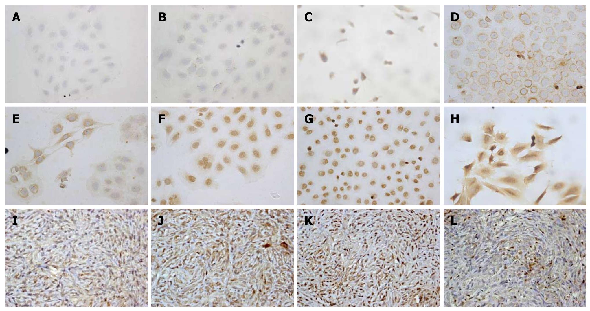
Figure 4 Immunohistochemical staining (× 400) analysis of Chang Gung CCA cells (upper two panels, A-H) and xenograft tissues of CCA mouse models (lower panel, I-L).
A: Negative control; B: Negative expression of K-ras; C: EGFR weakly expressed in a cytoplasmic distribution; D-G: Her-II, Biliary cytokeratin (CK19), COX-II, and Met diffusely expressed in a cytoplasmic distribution; H: MUC-4 strongly and diffusely expressed in a cytoplasmic and membranous distribution; I-L: The results revealed that CK-19, COX-II, Met, and MUC4 are diffusely expressed in a cytoplasmic distribution in the rat CCA xenograft.
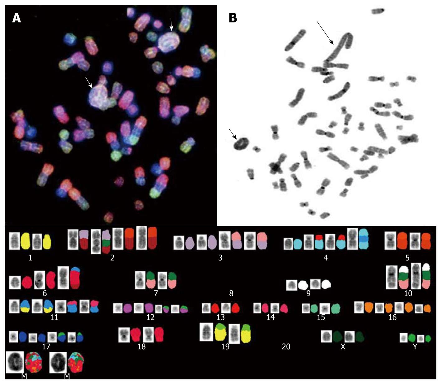
Figure 5 Examples of the karyotype spectral karyotyping analysis revealed by the 26th passage cell culture; the analysis established that, from the 25th week, TAA-induced CCA rat cells presented either (A) a ring chromosome (short arrows) or (B) giant rod chromosome (long arrow); complicated chromosomal alterations could be observed in most of the chromosomes and are illustrated at the bottom.
- Citation: Yeh CN, Lin KJ, Chen TW, Wu RC, Tsao LC, Chen YT, Weng WH, Chen MF. Characterization of a novel rat cholangiocarcinoma cell culture model-CGCCA. World J Gastroenterol 2011; 17(24): 2924-2932
- URL: https://www.wjgnet.com/1007-9327/full/v17/i24/2924.htm
- DOI: https://dx.doi.org/10.3748/wjg.v17.i24.2924


