Copyright
©2011 Baishideng Publishing Group Co.
World J Gastroenterol. Jun 7, 2011; 17(21): 2585-2591
Published online Jun 7, 2011. doi: 10.3748/wjg.v17.i21.2585
Published online Jun 7, 2011. doi: 10.3748/wjg.v17.i21.2585
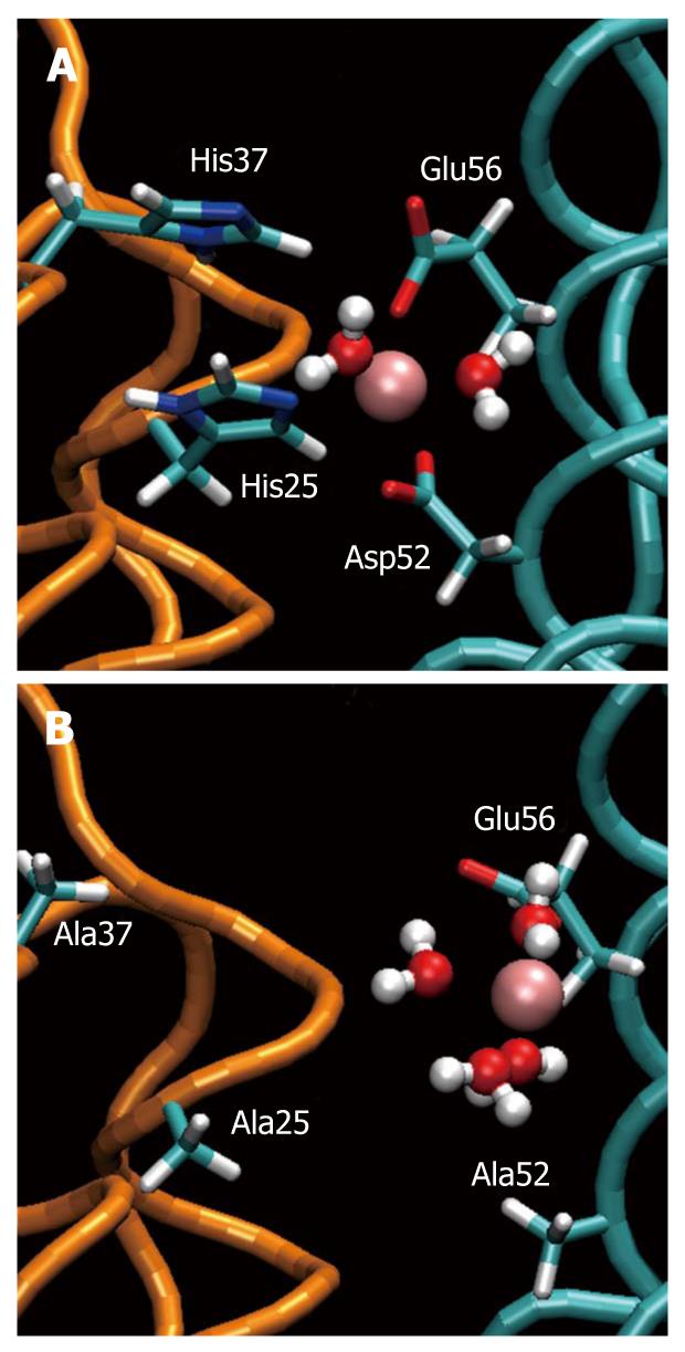
Figure 1 Ferroxidase site of Helicobacter pylori neutrophil activating protein.
A: The “ferroxidase site” in the equilibrated wild type. The iron ion (pink) is kept in position by Asp52, Glu56, His25 and His37. Two water molecules are attracted by Fe(II); B: The same site in the equilibrated mutant. The ferrous ion is attracted one-sidedly by Glu56 and Asp53 (not shown) loosing its ability to stabilize the dimer. Four water molecules are attracted by Fe(II).
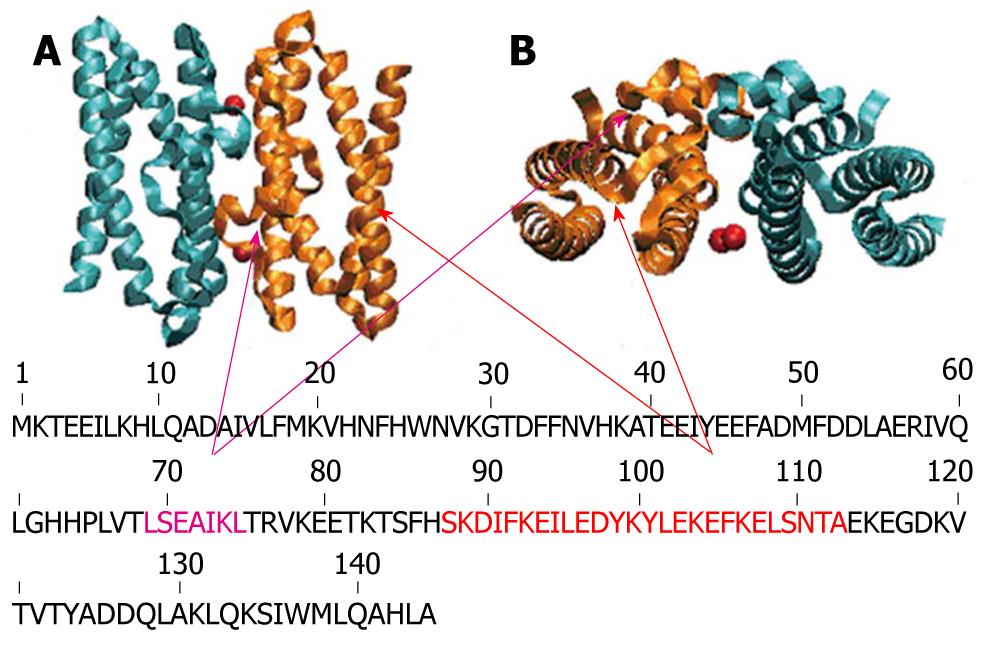
Figure 2 Schematic representation of exposed helices of Helicobacter pylori neutrophil activating protein.
Helicobacter pylori neutrophil activating protein dimer in stand up (A) and top view (B) with the exposed helices H3 and H4 (therefore possible candidates for interacting with the neutrophils) colored in violet and orange respectively.
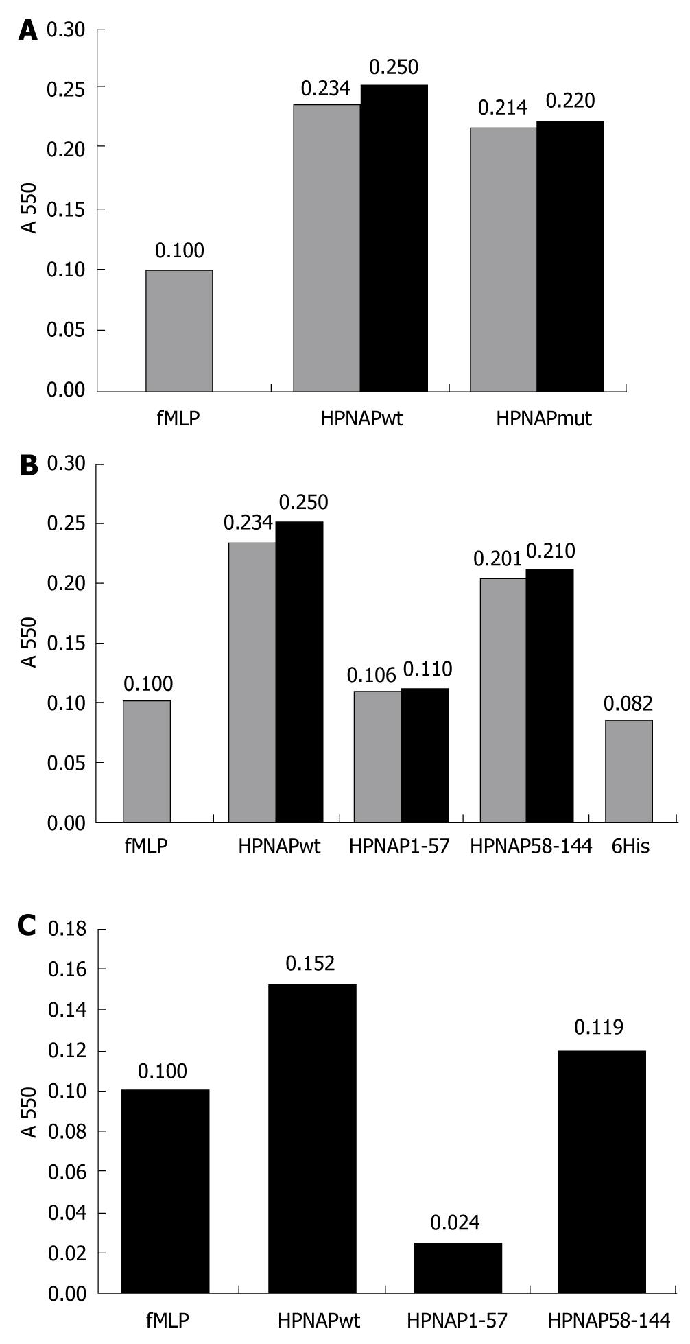
Figure 3 Neutrophil activation by Helicobacter pylori neutrophil activating protein.
A: Neutrophil activation by Helicobacter pylori neutrophil activating protein (HPNAP)-wt and HPNAPmut. Bar 1, activation by formyl-met-leu-pro peptide (fMLP) (control), bar 2, activation by HPNAPwt, bar 3, activation by HPNAPmut; B: Neutrophil activation by HPNAPwt-hexa-histidine peptide (6xHis), HPNAP1-57-6His, HPNAP58-144-6His and 6His peptide. Bar 1, activation by fMLP (control), bar 2, activation by HPNAPwt-6His, bar 3, activation by HPNAP1-57-6His, bar 4, activation by HPNAP58-144-6His, bar 5, activation by 6His peptide; C: Values after subtraction of “6His” value from these of “HPNAPwt”, “HPNAP1-57” and “HPNAP58-144”.
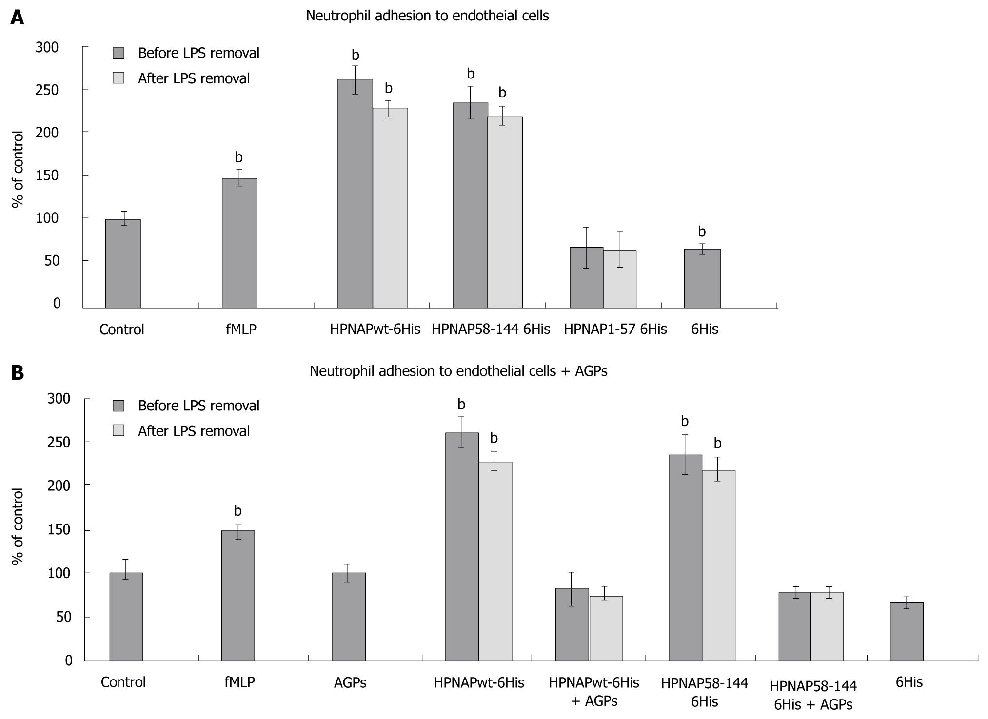
Figure 4 Neutrophil adhesion to endothelial cells.
A: Neutrophil adhesion to endothelial cells. Black bars indicate neutrophil adhesion to endothelial cells prior to lipopolysaccharides (LPS) removal while grey bars indicate the adhesion after LPS removal. The data represent triplicates from at least three independent experiments. Error bars indicate standard deviation (SD). Statistical evaluation was performed by Mann-Whitney test. Significant differences with the control values are marked by (bP < 0.001) bar 1, control, bar 2, formyl-met-leu-pro peptide (fMLP), positive control of the procedure, bar 3, Helicobacter pylori neutrophil activating protein (HPNAP)wt-hexa-histidine peptide (6xHis) effect on neutrophil adhesion to endothelial cells before and after LPS removal, bar 4, HPNAP58-144-6xHis effect before and after LPS removal, bar 5, HPNAP1-57-6xHis effect before and after LPS removal, bar 6, 6xHis effect; B: Effect of arabino galactan proteins (AGPs) on neutrophil adhesion to endothelial cells, bar 1, control, bar 2, fMLP, positive control of the procedure, bar 3, effect of AGPs, bar 4, HPNAPwt-6xHis effect before and after LPS removal, bar 5, HPNAPwt-6xHis and AGPs co-effect, before and after LPS removal, bar 6, HPNAP58-144-6xHis effect before and after LPS removal, bar 7, HPNAP58-144-6xHis and mastic gum extract co-effect, before and after LPS removal, bar 8, 6xHis effect.
-
Citation: Choli-Papadopoulou T, Kottakis F, Papadopoulos G, Pendas S.
Helicobacter pylori neutrophil activating protein as target for new drugs againstH. pylori inflammation. World J Gastroenterol 2011; 17(21): 2585-2591 - URL: https://www.wjgnet.com/1007-9327/full/v17/i21/2585.htm
- DOI: https://dx.doi.org/10.3748/wjg.v17.i21.2585


