Copyright
©2011 Baishideng Publishing Group Co.
World J Gastroenterol. May 21, 2011; 17(19): 2446-2449
Published online May 21, 2011. doi: 10.3748/wjg.v17.i19.2446
Published online May 21, 2011. doi: 10.3748/wjg.v17.i19.2446
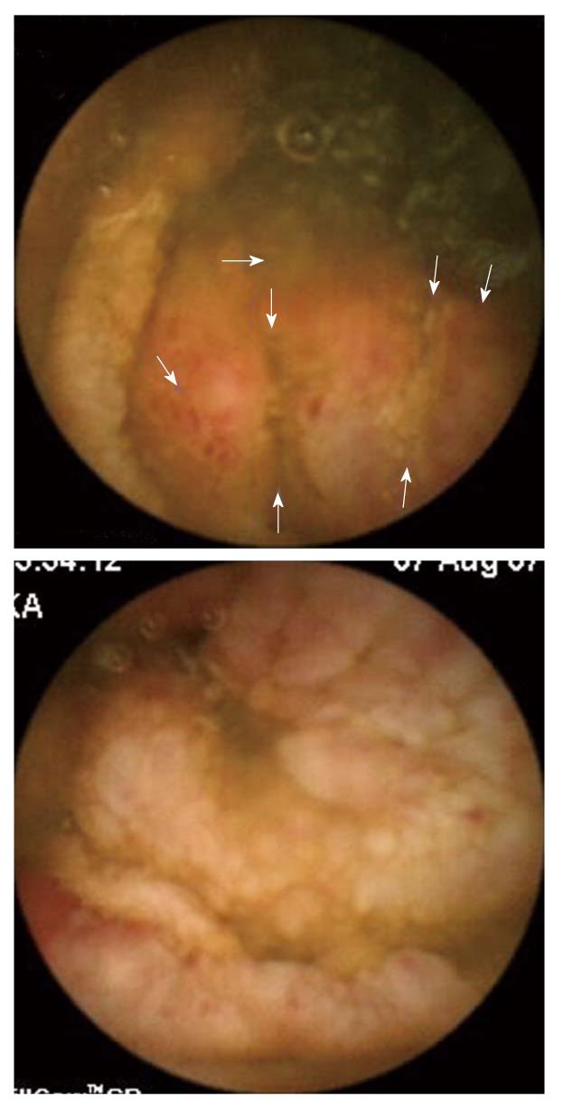
Figure 1 Capsule images of ulcerated, lumpy mucosa from the distal jejunum to proximal ileum.
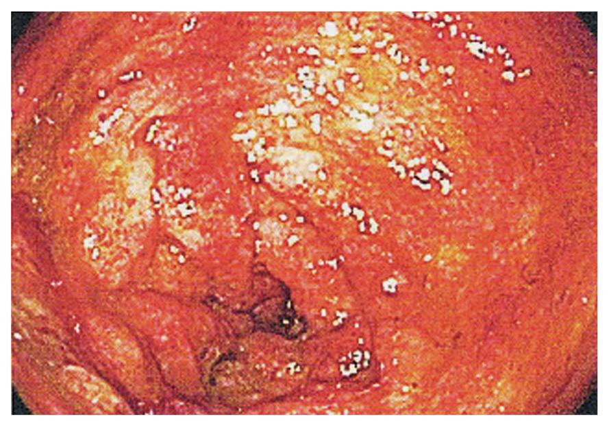
Figure 2 Colonoscopy image of erythematous, ulcerated, lumpy mucosa of the terminal ileum.
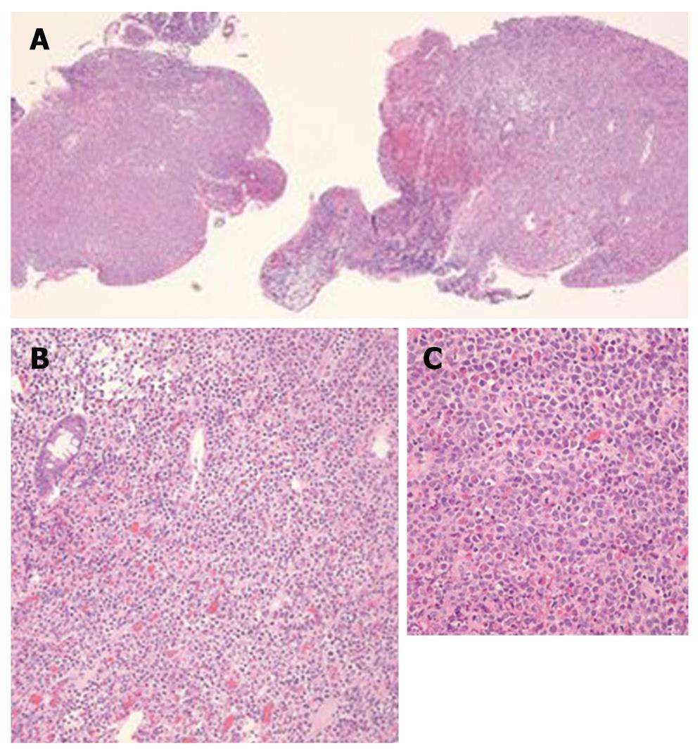
Figure 3 Biopsies from the terminal ileum showing extramedullary myeloid cell tumor (granulocytic sarcoma).
A: 40 x magnification; B: 200 x magnification; C: 400 x magnification.
- Citation: Kwan LY, Targan SR, Shih DQ. A case of steroid-dependent myeloid granulocytic sarcoma masquerading as Crohn’s disease. World J Gastroenterol 2011; 17(19): 2446-2449
- URL: https://www.wjgnet.com/1007-9327/full/v17/i19/2446.htm
- DOI: https://dx.doi.org/10.3748/wjg.v17.i19.2446


