Copyright
©2011 Baishideng Publishing Group Co.
World J Gastroenterol. May 7, 2011; 17(17): 2199-2205
Published online May 7, 2011. doi: 10.3748/wjg.v17.i17.2199
Published online May 7, 2011. doi: 10.3748/wjg.v17.i17.2199
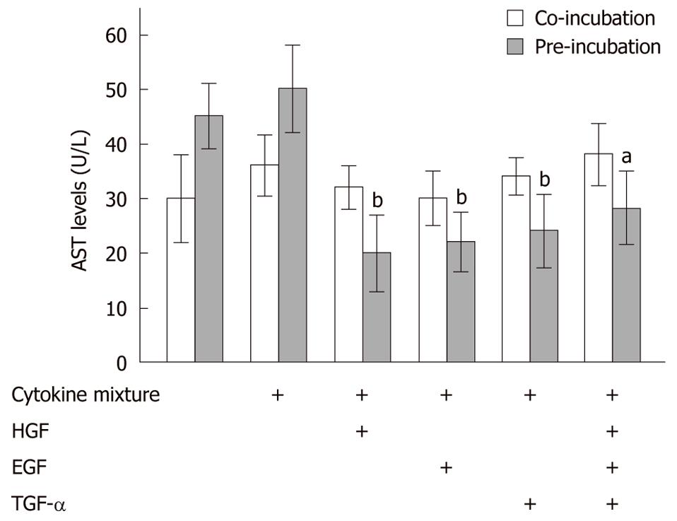
Figure 1 Pre-stimulation with growth factors significantly reduces cellular damage induced by lipopolysaccharide-containing cytokine mixture.
Primary rat hepatocytes (N = 5, n = 3) treated with lipopolysaccharide (LPS)-containing cytokine mixture (CM) for 24 h showed a slight increase in aspartate aminotransferase (AST) leakage into the culture supernatant. Co-incubation with the hepatocyte growth factor (HGF), epidermal growth factor (EGF) and transforming growth factor (TGF)-α, individually or in combination, did not reduce AST leakage significantly (empty bars). However, preincubation with these hepatotropic growth factors resulted in a significant reduction in AST leakage (grey bars). aP < 0.05, bP < 0.005 vs corresponding rat hepatocytes treated with LPS-containing CM alone.
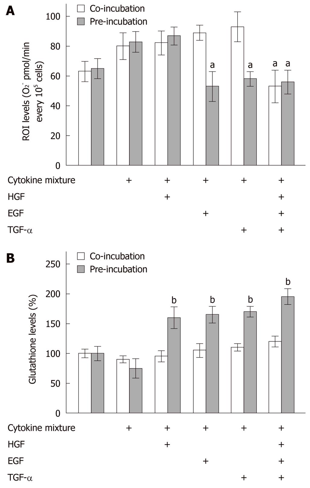
Figure 2 Increased oxidative stress in rat hepatocytes treated with lipopolysaccharide-containing cytokine mixture.
A: Treatment of primary rat hepatocytes (N = 5, n = 3) with lipopolysaccharide (LPS)-containing cytokine mixture (CM) for 24 h caused a slight increase in O2- production. B: Cellular glutathione levels were not significantly affected by this treatment. Co-incubation with single hepatocyte growth factor (HGF), epidermal growth factor (EGF) or transforming growth factor (TGF)-α did not reduce reactive oxygen intermediate (ROI) production significantly. Co-incubation with the hepatotropic growth factor mixture alone was able to reduce ROI production significantly. Furthermore, co-incubation with the growth factors did not alter cellular glutathione levels (empty bars). On the other hand, preincubation with these growth factors, individually or in combination, significantly reduced ROI production (except for pretreatment with HGF alone). Preincubation with the different growth factors increased cellular glutathione significantly (grey bars). aP < 0.05, bP < 0.001 vs corresponding rat hepatocytes treated with LPS-containing CM alone.
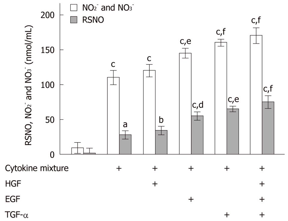
Figure 3 Hepatotropic growth factors increase nitric oxide formation in primary rat hepatocytes.
NO2- plus NO3- (empty bars) and RSNO (grey bars) levels demonstrated a significant increase after hepatocyte (N = 5, n = 3) pretreatment with growth factors and subsequent stimulation with lipopolysaccharide (LPS)-containing cytokine mixture (CM), if compared to treatment with LPS-containing CM alone [except for pretreatment with hepatocyte growth factor (HGF) alone]. aP < 0.05, bP < 0.005, cP < 0.001 vs corresponding untreated rat hepatocytes; dP < 0.05, eP < 0.005, fP < 0.001 vs corresponding rat hepatocytes treated with LPS-containing CM alone. NO: Nitric oxide; EGF: Epidermal growth factor; TGF: Transforming growth factor.
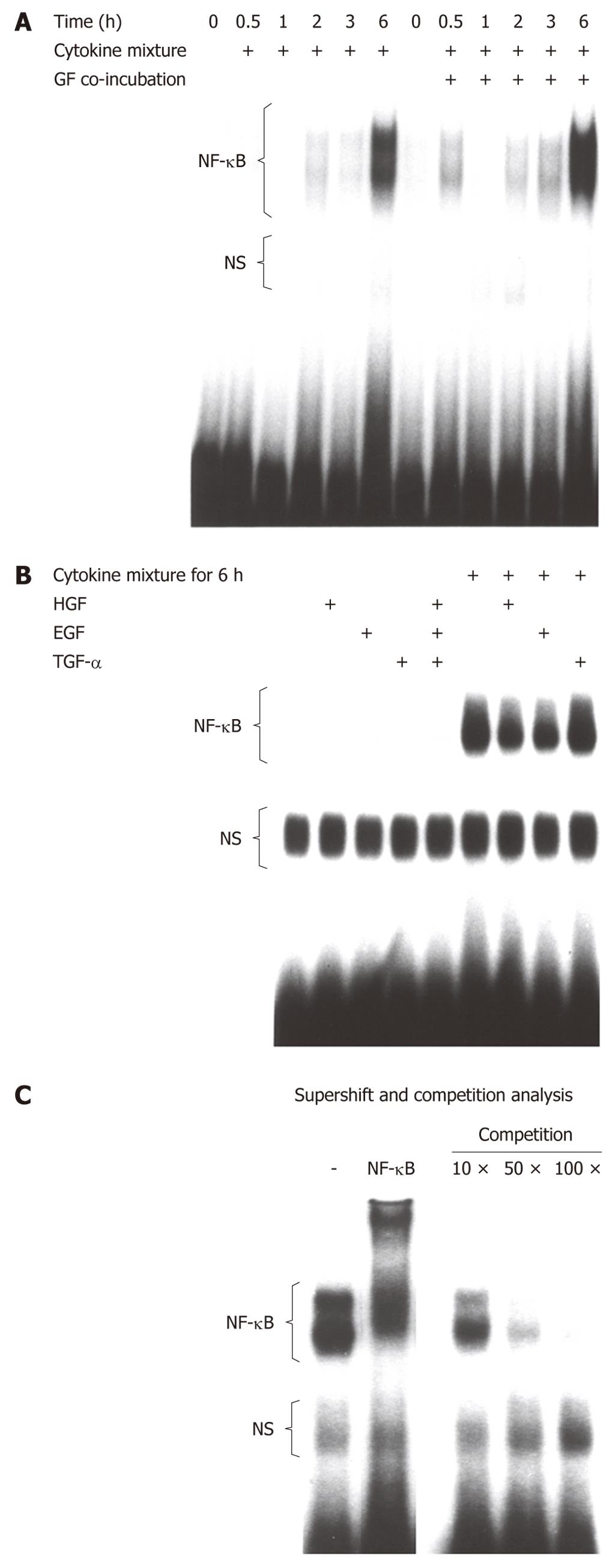
Figure 4 No additional nuclear factor-κB activation by hepatotropic growth factors.
Electrophoretic mobility shift assay for nuclear factor (NF)-κB after hepatocyte pretreatment with hepatotropic growth factors and subsequent stimulation with lipopolysaccharide (LPS)-containing cytokine mixture (CM) demonstrated that stimulation with growth factors combined (A) or individually (B) did not further increase NF-κB expression when compared to stimulation with LPS-containing CM alone. Super shift and competition analysis (C) of NF-κB. HGF: Hepatocyte growth factor; EGF: Epidermal growth factor; TGF: Transforming growth factor.
- Citation: Glanemann M, Knobeloch D, Ehnert S, Culmes M, Seeliger C, Seehofer D, Nussler AK. Hepatotropic growth factors protect hepatocytes during inflammation by upregulation of antioxidative systems. World J Gastroenterol 2011; 17(17): 2199-2205
- URL: https://www.wjgnet.com/1007-9327/full/v17/i17/2199.htm
- DOI: https://dx.doi.org/10.3748/wjg.v17.i17.2199


