Copyright
©2011 Baishideng Publishing Group Co.
World J Gastroenterol. May 7, 2011; 17(17): 2172-2177
Published online May 7, 2011. doi: 10.3748/wjg.v17.i17.2172
Published online May 7, 2011. doi: 10.3748/wjg.v17.i17.2172
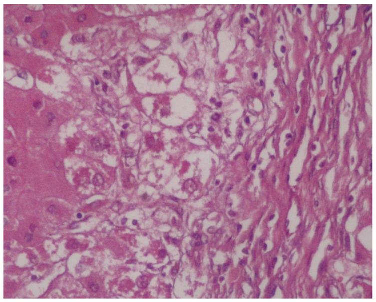
Figure 1 Zone 1 distribution of Mallory-Denk Bodies in primary biliary cirrhosis (× 400).
This distribution was observed in both early and later stages of primary biliary cirrhosis, such as cirrhosis.
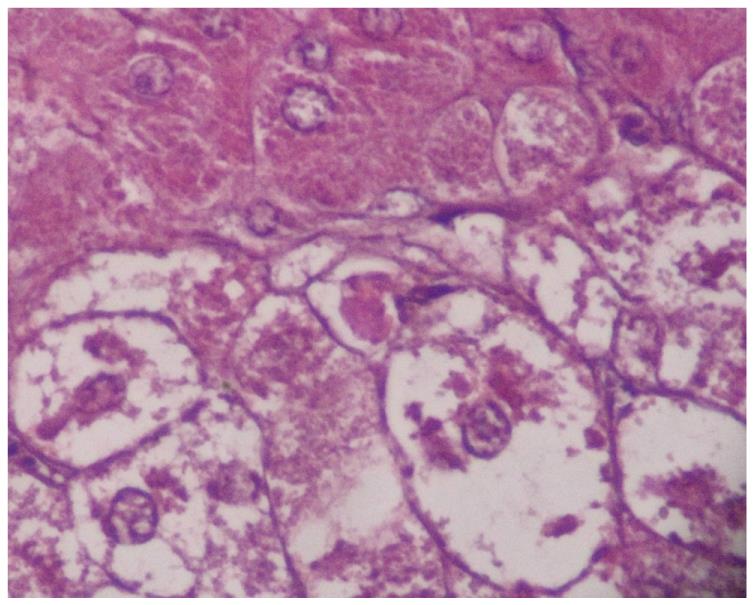
Figure 2 Mallory-Denk Bodies, located perinuclear, has been shown as a larger picture in a patient with primary biliary cirrhosis (× 1000).
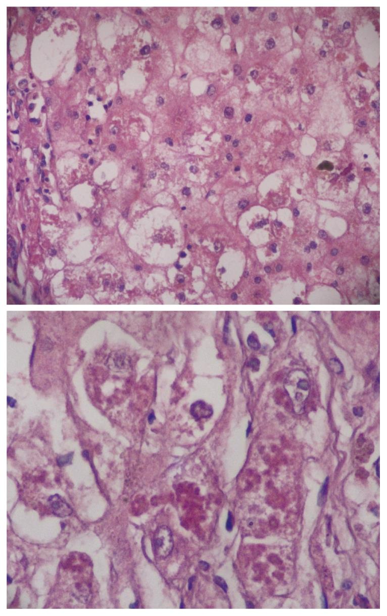
Figure 3 Mallory-Denk Bodies, located both perinuclear and intracytoplasmic, was observed in zone 1 in early stages of Wilson disease, but in every zone at the later stages, such as cirrhosis (× 400, 1000, respectively).
Hydrophobic degeneration was more exaggerated. The diameter of Mallory-Denk Bodies was not homogenous in patients with Wilson disease.
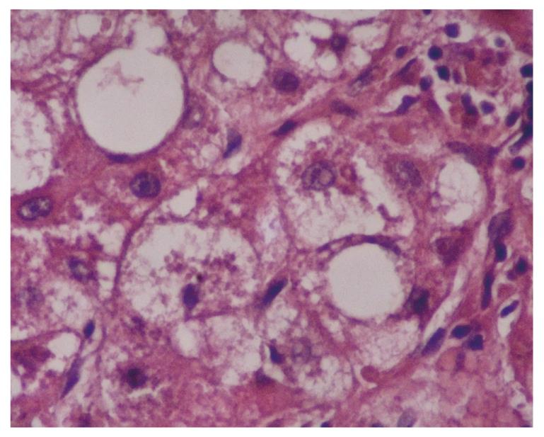
Figure 4 Perinuclear location of Mallory-Denk Bodies in patients with hepatocellular carcinoma depends on the cause (× 1000).
In this case, hepatitis B is responsible from Mallory-Denk Bodies.
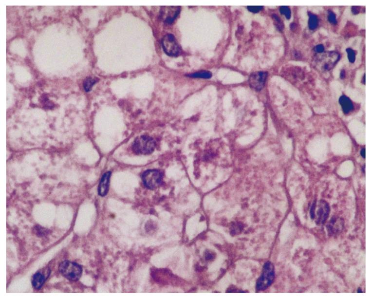
Figure 5 Tiny and uniform Mallory-Denk Bodies was seen in patients with nonalcoholic steatohepatitis (× 1000).
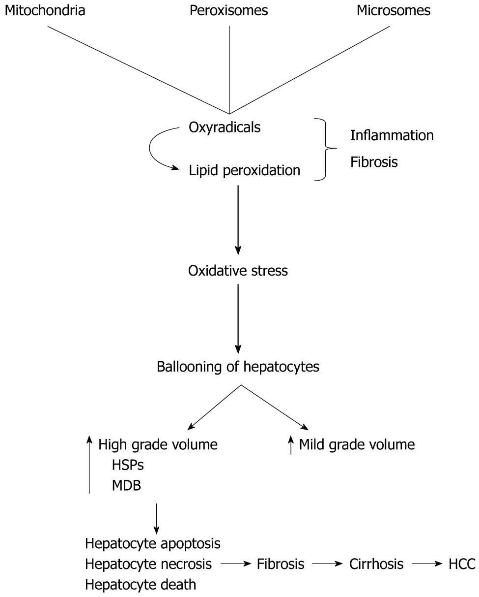
Figure 6 Shows development of Mallory-Denk Bodies in ballooned hepatocytes.
MDB: Mallory-Denk Bodies; HSPs: Heat shock proteins; HCC: Hepatocellular carcinoma.
- Citation: Basaranoglu M, Turhan N, Sonsuz A, Basaranoglu G. Mallory-Denk Bodies in chronic hepatitis. World J Gastroenterol 2011; 17(17): 2172-2177
- URL: https://www.wjgnet.com/1007-9327/full/v17/i17/2172.htm
- DOI: https://dx.doi.org/10.3748/wjg.v17.i17.2172


