Copyright
©2011 Baishideng Publishing Group Co.
World J Gastroenterol. Apr 21, 2011; 17(15): 2054-2057
Published online Apr 21, 2011. doi: 10.3748/wjg.v17.i15.2054
Published online Apr 21, 2011. doi: 10.3748/wjg.v17.i15.2054
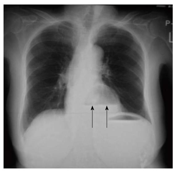
Figure 1 A chest X-ray film at admission, revealing air in the dilated stomach associated with air-fluid level (arrows), which was dislocated into the chest.
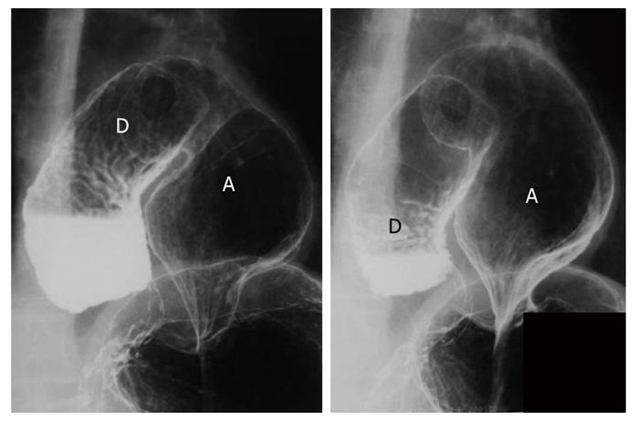
Figure 2 An upper gastrointestinal series performed preoperatively, revealing dislocated distal portion of the stomach (A) and duodenum (D) into the thoracic cavity rotated in the mesenteric axis, which corresponded to a mesenterioaxial volvulus of the upper gastrointestinal tract.
Figure 3 Abdominal computed tomography scan.
An incarcerated upper gastrointestinal tract in the thoracic cavity (arrows). The gastrointestinal tract dislocated in the thoracic cavity was significantly dilated, where fluid had accumulated.
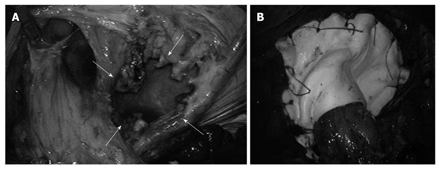
Figure 4 Laparoscopic view after initial reduction of the stomach from the chest into the abdomen.
Sliding hernia and parahiatal hernia. A: A paraesophageal aperture (arrows) was large-sized and was located at the left side of the left crus of the esophageal hiatus; B: Hernia orifice was closed using the mesh reinforcement.
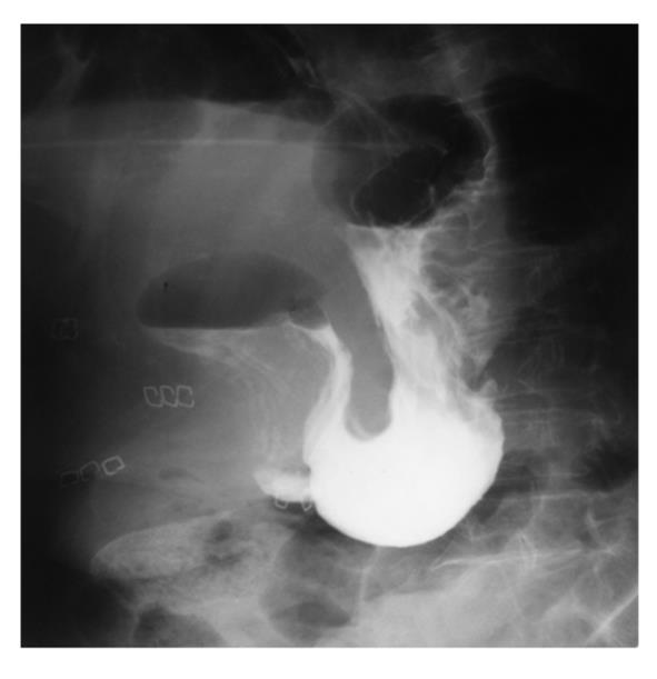
Figure 5 A postoperative upper gastrointestinal series performed at 5 d after surgery, showing the normal intraperitoneal location of the stomach and duodenum.
- Citation: Inaba K, Sakurai Y, Isogaki J, Komori Y, Uyama I. Laparoscopic repair of hiatal hernia with mesenterioaxial volvulus of the stomach. World J Gastroenterol 2011; 17(15): 2054-2057
- URL: https://www.wjgnet.com/1007-9327/full/v17/i15/2054.htm
- DOI: https://dx.doi.org/10.3748/wjg.v17.i15.2054


