Copyright
©2011 Baishideng Publishing Group Co.
World J Gastroenterol. Apr 7, 2011; 17(13): 1787-1790
Published online Apr 7, 2011. doi: 10.3748/wjg.v17.i13.1787
Published online Apr 7, 2011. doi: 10.3748/wjg.v17.i13.1787
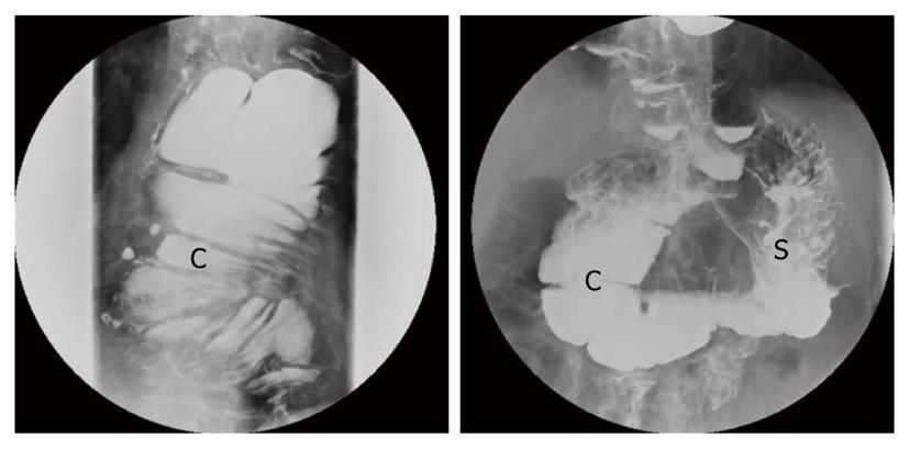
Figure 1 Esophagography showing the interposition of the colon (C) and the gastric remnant (S).
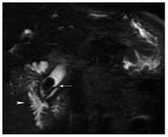
Figure 2 Magnetic resonance cholangiopancreatography showing a stone in the distal common bile duct (arrow).
The arrowhead shows the second portion of the duodenum.
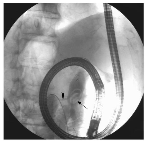
Figure 3 The radio-opaque standard guidewire (arrowhead) was inserted through the working channel of the gastroscope.
Figure 4 With the patient in the left-lateral position, endoscopic retrograde cholangiopancreatography showed a filling defect in the distal part of the common bile duct (arrow).
The arrowhead shows the pancreatic duct.
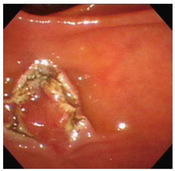
Figure 5 Complete sphincterotomy.
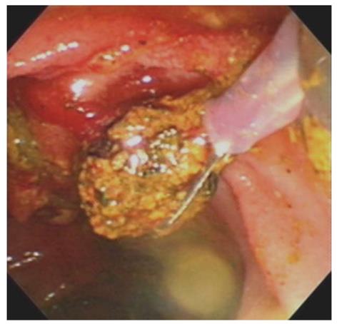
Figure 6 The pigment stone retrieved by a Dormia basket.
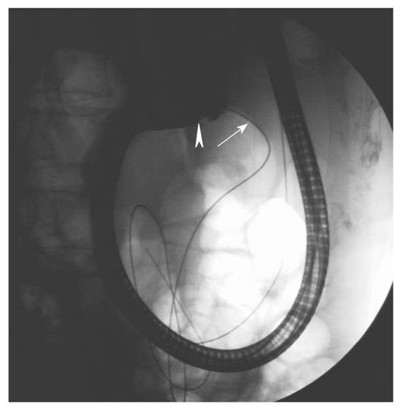
Figure 7 The duodenoscope (arrowhead) was pushed along the guidewire (arrow) at the same axis under fluoroscopic guidance .
- Citation: Yii CY, Chou JW, Peng YC, Chow WK. Application of a wire-guided side-viewing duodenoscope in total esophagectomy with colonic interposition. World J Gastroenterol 2011; 17(13): 1787-1790
- URL: https://www.wjgnet.com/1007-9327/full/v17/i13/1787.htm
- DOI: https://dx.doi.org/10.3748/wjg.v17.i13.1787


