Copyright
©2011 Baishideng Publishing Group Co.
World J Gastroenterol. Apr 7, 2011; 17(13): 1732-1738
Published online Apr 7, 2011. doi: 10.3748/wjg.v17.i13.1732
Published online Apr 7, 2011. doi: 10.3748/wjg.v17.i13.1732
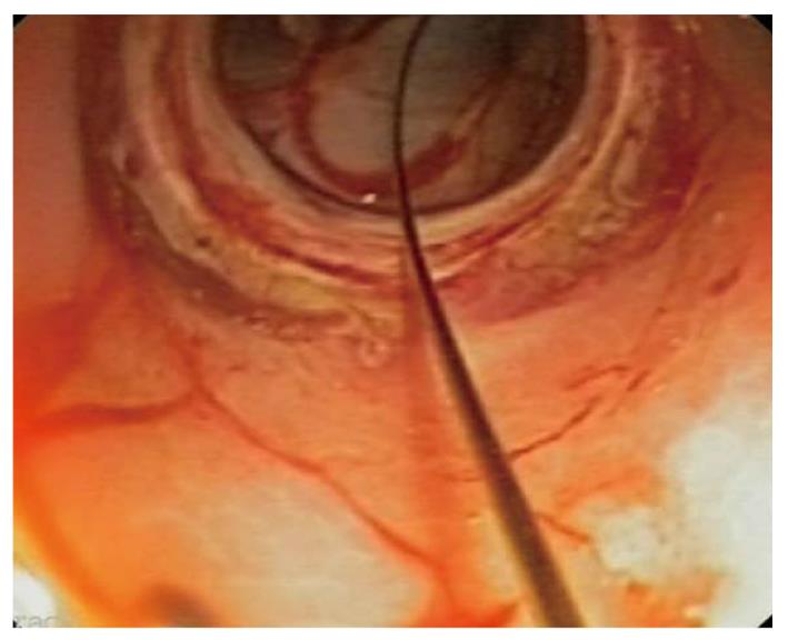
Figure 1 The incision is enlarged with a balloon dilator.
Through the balloon we can see peritoneal structures.
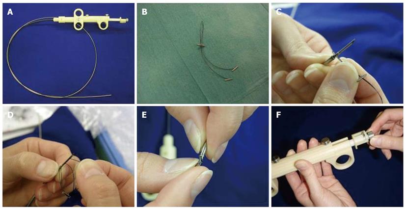
Figure 2 Description of the tissue anchoring device.
A: Single 18-gauge flexible needle catheter; B: Bifurcated nylon thread (“Y” shaped) with 3 small tags (2 regular tags fixed at both bifurcated distal ends and the other tag stopper at the single proximal end, which is used for tightening); C: The proximal end of the thread is fixed to the needle with a small metallic guide; D: The two distal tags are consecutively inserted into the needle; E: The needle is pulled back into the sheath inserting also the stopper tag; F: The pusher button allows releasing one tag at each side of the incision.
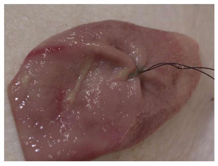
Figure 3 The incision looks completely sealed after the insertion of one brace-bar and the stomach is able to maintain air distension.
- Citation: Guarner-Argente C, Córdova H, Martínez-Pallí G, Navarro-Ripoll R, Rodríguez-d’Jesús A, Miguel CR, Beltrán M, Fernández-Esparrach G. Gastrotomy closure with a new tissue anchoring device: A porcine survival study. World J Gastroenterol 2011; 17(13): 1732-1738
- URL: https://www.wjgnet.com/1007-9327/full/v17/i13/1732.htm
- DOI: https://dx.doi.org/10.3748/wjg.v17.i13.1732


