Copyright
©2011 Baishideng Publishing Group Co.
World J Gastroenterol. Mar 28, 2011; 17(12): 1584-1593
Published online Mar 28, 2011. doi: 10.3748/wjg.v17.i12.1584
Published online Mar 28, 2011. doi: 10.3748/wjg.v17.i12.1584
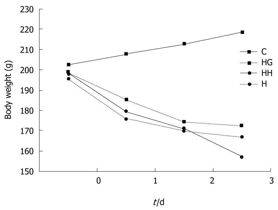
Figure 1 Body weight changes in rats of different groups.
The body weight of rats in group C increased stably, and reduced body weight was observed in all rats exposed to hypobaric hypoxia. Decreased body weight was most evident in group HH, followed by groups H and HG. C: Control group; H: Hypobaric hypoxia group; HH: Hypobaric hypoxia plus starvation group; HG: Hypobaric hypoxia plus Gln group.
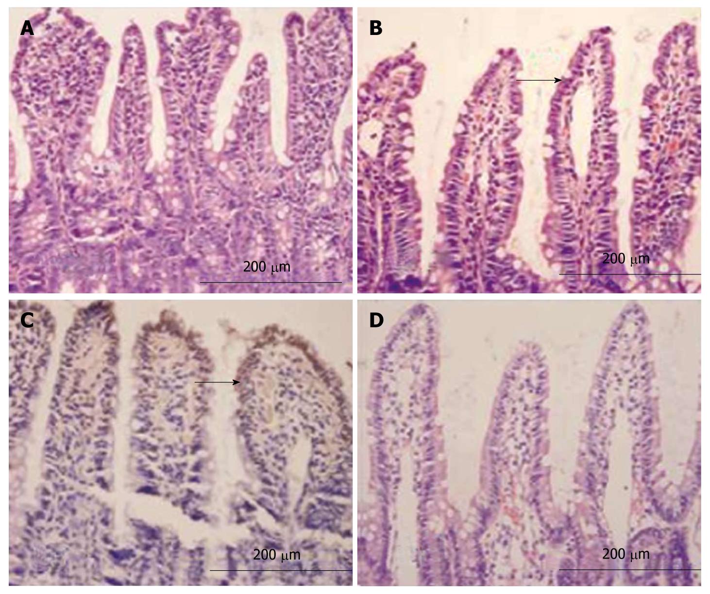
Figure 2 Light microscopy.
Smooth intestinal mucosa with intact epithelia and ordered villi in group C (A), exfoliated and incomplete intestinal mucosa with thickened mucosa and reduced villi accompanying irregular morphology and disorganized villous epithelia in group H (B), atrophic and thinned villi accompanying a loose and disordered arrangement as well as edema and infiltration of inflammatory cells in mastoid lamina of villi and lodged and exfoliated villi with loss of goblet cells and red blood cell effusion around the capillaries in group HH (C), relatively intact intestinal mucosal villi with ordered arrangement and alleviated edema in mastoid lamina of villi accompanying a few infiltrated inflammatory cells in group HG (D) (HE, × 200).
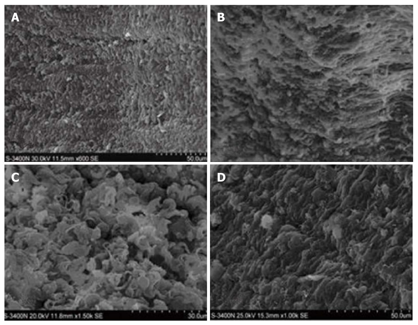
Figure 3 Scanning electron microscopy.
A smooth surface of intestinal mucosa with a clear structure as well as complete and orderly villi in group C (A) (× 6900), atrophic epithelial structure of intestinal mucosa and lodging villi accompanying a rough surface with disordered villi and widened villous spaces in group H (B) (× 6900), severely injured intestinal mucosa as well as disc-shaped cells and cellulose in mucosal defects along with evident atrophy and disordered villi with widened villous spaces and exfoliated microvilli in group HH (C) (× 11 500), almost intact intestinal mucosa with orderly villi and less effusion but no disc-shaped cells and cellulose in group HG (D) (× 11 500).
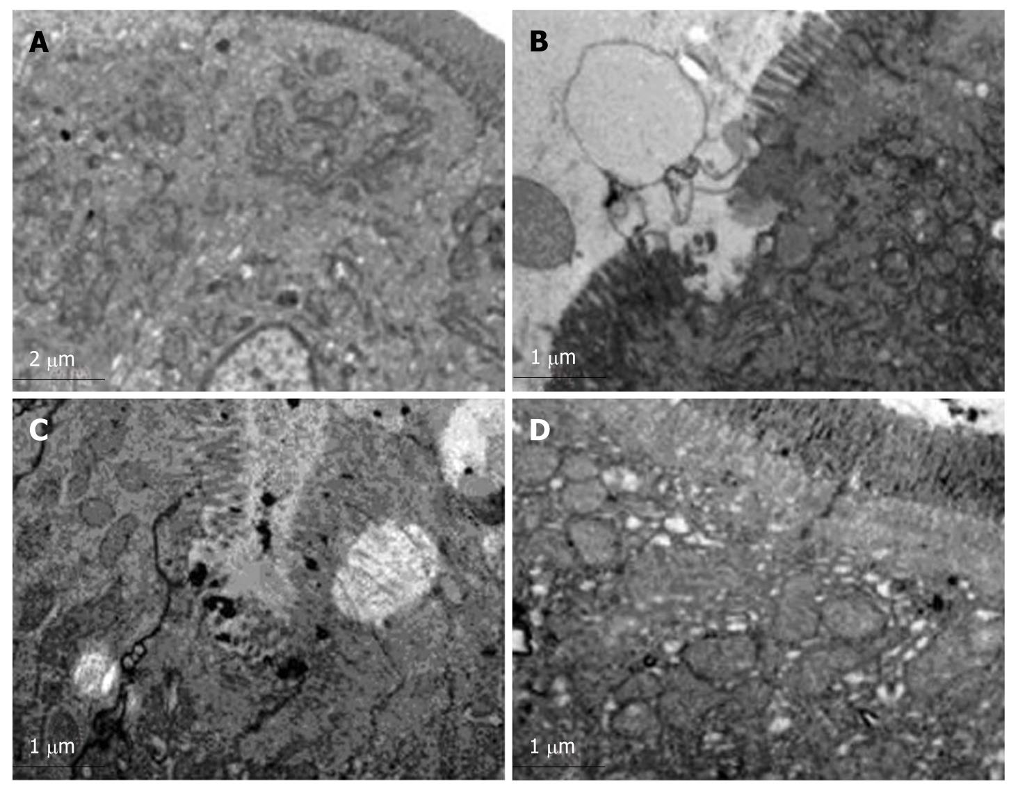
Figure 4 Transmission electron microscopy with nitric acid lanthanum tagging.
Orderly mucosal villi and integrated tight junctions as well as intact organelles with regular nuclei in group C (A), exfoliated and incomplete microvilli accompanying widened intercellular spaces as well as swollen endoplasmic reticulum and mitochondria and a small number of lanthanum granules in tissue spaces in group H (B), dilated Golgi complex with irregular nuclei and edge aggregation in chromatin as well as lanthanum granules in the tight junction gap and cells in group HH (C), mildly deformed microvilli and swollen mitochondria in lamina propria accompanying a small number of lanthanum granules confined to vessels and epithelial surface in group HG (D) (× 8900).
- Citation: Zhou QQ, Yang DZ, Luo YJ, Li SZ, Liu FY, Wang GS. Over-starvation aggravates intestinal injury and promotes bacterial and endotoxin translocation under high-altitude hypoxic environment. World J Gastroenterol 2011; 17(12): 1584-1593
- URL: https://www.wjgnet.com/1007-9327/full/v17/i12/1584.htm
- DOI: https://dx.doi.org/10.3748/wjg.v17.i12.1584


