Copyright
©2011 Baishideng Publishing Group Co.
World J Gastroenterol. Mar 21, 2011; 17(11): 1494-1500
Published online Mar 21, 2011. doi: 10.3748/wjg.v17.i11.1494
Published online Mar 21, 2011. doi: 10.3748/wjg.v17.i11.1494
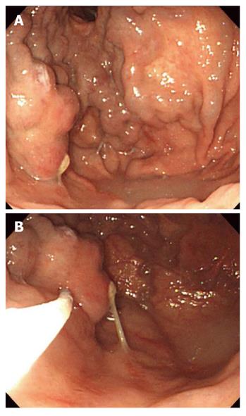
Figure 1 Endoscopic Histoacryl® injection for treatment of gastric varices.
A: Endoscopic image showing a large gastric varix; B: Injection of Histoacryl® into the gastric varix using the catheter.
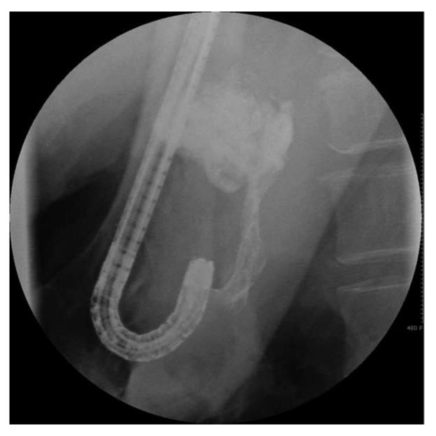
Figure 2 X-ray showing lipiodol in the gastric varix during the procedure.
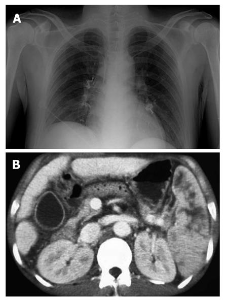
Figure 3 Embolic complications of Histoacryl® injection.
A: Chest radiography showing pulmonary embolism after Histoacryl® injection; B: Computed tomography showing the presence of lipiodol in the splenic vein after Histoacryl® injection.
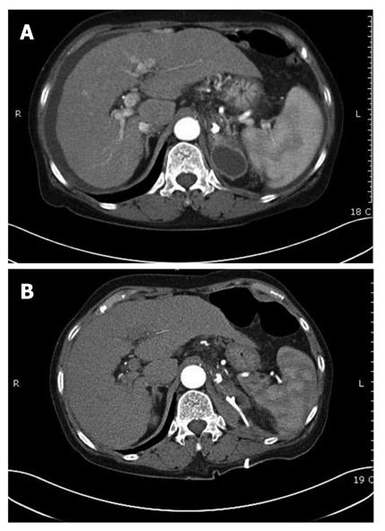
Figure 4 Adrenal abscess after Histoacryl® injection.
A: Computed tomography scan showing adrenal abscess after Histoacryl® injection; B: Adrenal abscess was resolved after PCD insertion.
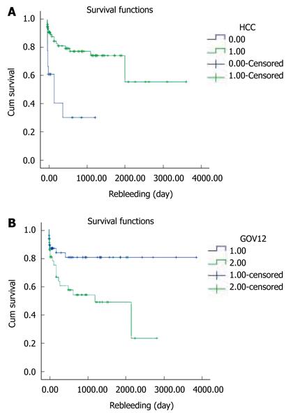
Figure 5 The 1-year rebleeding rate using the Kaplan-Meier method and then log rank test.
A: Comparing the groups with hepatocellular carcinoma (HCC) or without HCC; B: Comparing GOV1 and GOV2. GOV: Gastro-esophageal varices.
Figure 6 Survival of Histoacryl® injected patients.
A: Survival of 127 Histoacryl®-injected patients using the Kaplan-Meier method. The cumulative survival rates of the 127 patients at 6 mo, and 1, 3 and 5 years were 92.1%, 84.2%, 64.2%, and 45.3%, respectively; B: Survival of the 27 patients who underwent primary prophylaxis, using the Kaplan-Meier method. The 6 mo cumulative survival rate of these patients was 75%.
- Citation: Kang EJ, Jeong SW, Jang JY, Cho JY, Lee SH, Kim HG, Kim SG, Kim YS, Cheon YK, Cho YD, Kim HS, Kim BS. Long-term result of endoscopic Histoacryl® (N-butyl-2-cyanoacrylate) injection for treatment of gastric varices. World J Gastroenterol 2011; 17(11): 1494-1500
- URL: https://www.wjgnet.com/1007-9327/full/v17/i11/1494.htm
- DOI: https://dx.doi.org/10.3748/wjg.v17.i11.1494


