Copyright
©2011 Baishideng Publishing Group Co.
World J Gastroenterol. Jan 7, 2011; 17(1): 21-27
Published online Jan 7, 2011. doi: 10.3748/wjg.v17.i1.21
Published online Jan 7, 2011. doi: 10.3748/wjg.v17.i1.21
Figure 1 Esophageal surface epithelium visualized using intravenous fluorescein.
A: Capillary loops (arrows) of the esophageal papillae; B: Dilatation of intercellular spaces and increased vasculature off the papillae in reflux esophagitis; C: Presence of cylindrical epithelial cell and goblet cells (arrows) in the distal esophagus suggesting Barrett’s epithelium.
Figure 2 Confocal laser endomicroscopy of the stomach using intravenous fluorescein.
A: Columnar epithelium of the antral gastric mucosa and regulated microvascular matrix; B: Atrophic gastritis with intestinal metaplasia and presence of goblet cells (arrows); C: Early gastric cancer with disorganized tissue architecture, few regular crypts (arrowhead) and very tortuous, dilated, irregular vessels (arrows).
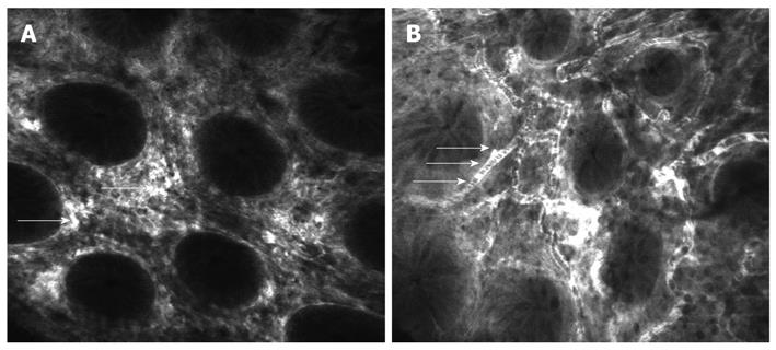
Figure 3 Confocal laser endomicroscopy of the normal colon using intravenous fluorescein.
A: Normal aspect of colonic mucosa showing regular architecture of crypts and capillaries of lamina propria (arrows); B: Dark shadows in the lumen of the vessels representing the red blood cells (arrows).
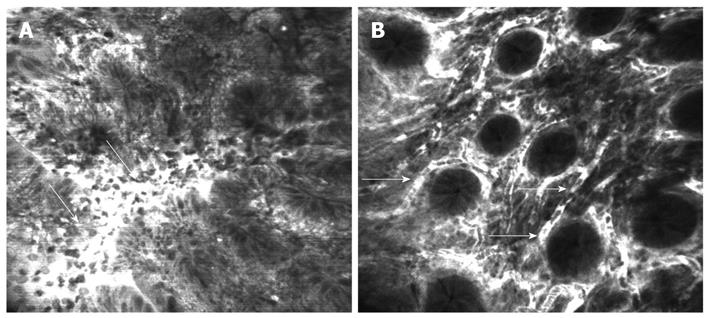
Figure 4 Confocal laser endomicroscopy of the colon using intravenous fluorescein.
A: Colon carcinoma with total disorganization of cell architecture, invasion and destruction of the vessels with leakage of fluorescein (arrows); B: Severe inflammatory changes in ulcerative colitis with cellular infiltrate causing an increase in the distance between crypts and excessive vascularity (arrows).
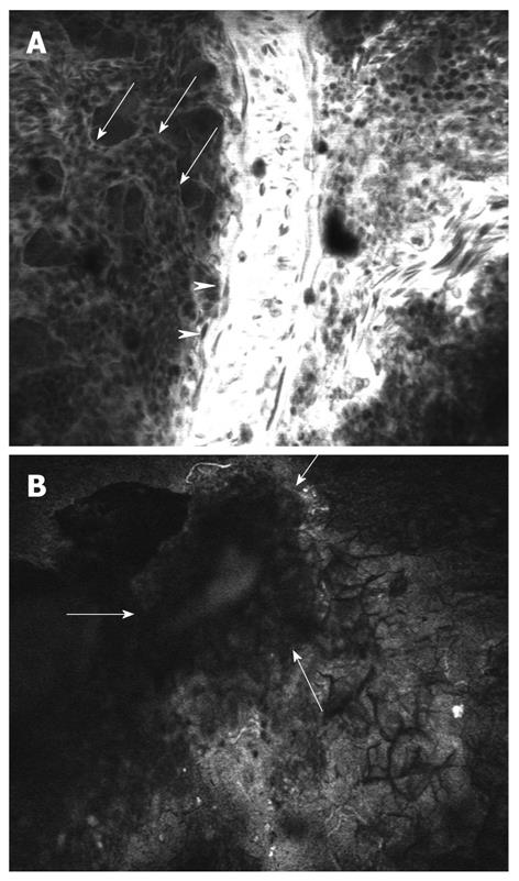
Figure 5 Confocal laser microscopy of the chick embryo chorioallantoic membrane.
A: Chick normal chorioallantoic membrane with visualization of large vessels (arrowheads), medium vessels (arrows) and circulating nucleated erythrocytes; B: Fragment of viable human colon cancer tissue (arrows) implanted on chick embryo chorioallantoic membrane.
- Citation: Gheonea DI, Cârţână T, Ciurea T, Popescu C, Bădărău A, Săftoiu A. Confocal laser endomicroscopy and immunoendoscopy for real-time assessment of vascularization in gastrointestinal malignancies. World J Gastroenterol 2011; 17(1): 21-27
- URL: https://www.wjgnet.com/1007-9327/full/v17/i1/21.htm
- DOI: https://dx.doi.org/10.3748/wjg.v17.i1.21


