Copyright
©2011 Baishideng Publishing Group Co.
World J Gastroenterol. Jan 7, 2011; 17(1): 111-117
Published online Jan 7, 2011. doi: 10.3748/wjg.v17.i1.111
Published online Jan 7, 2011. doi: 10.3748/wjg.v17.i1.111
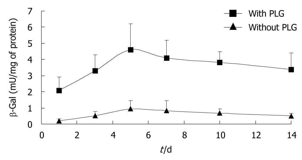
Figure 1 Time-course study of the effect of the poly-L-glutamate formulation on transgene expression of pLac in the tibialis anterior muscle after electroporation transfer.
The plasmids were transferred with (square) and without (triangle) poly-L-glutamate (PLG) formulation. The values shown are mean ± SD, n = 8.
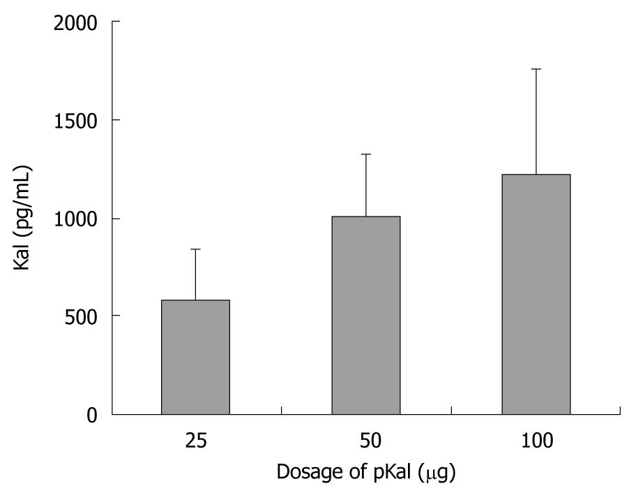
Figure 2 Dose-dependent kallistatin concentrations in the serum responded to intramuscular electrotransfer of a plasmid pKal in poly-L-glutamate formulation.
The values shown are mean ± SD, n = 8. Kal: Kallistatin.
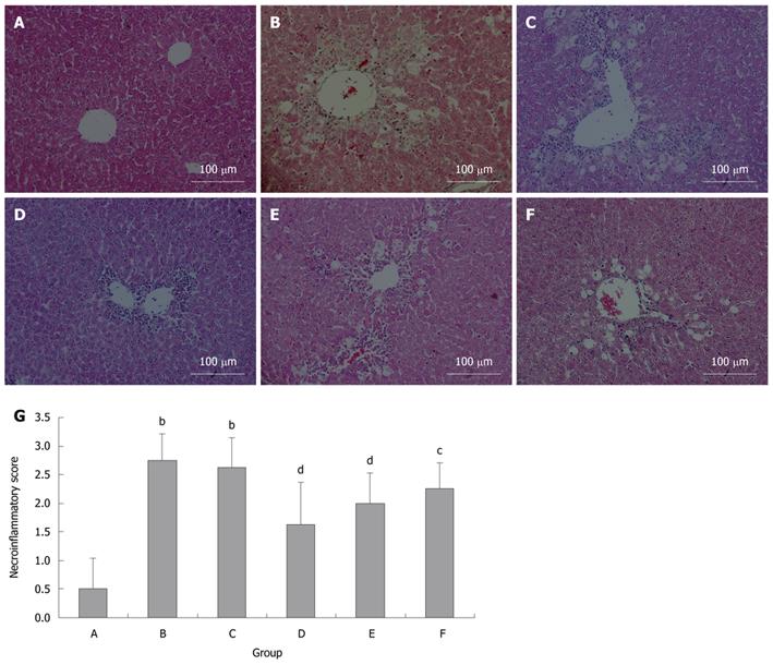
Figure 3 Protective effects of kallistatin expression against carbon tetrachloride-induced liver injury in mice.
HE staining was performed on paraffin embedded sections of the liver tissues. Representative sections are shown for each group. A: Control; B: Carbon tetrachloride (CCl4); C: pLac + CCl4; D: pKal (100 μg) + CCl4; E: pKal (50 μg) + CCl4; F: pKal (25 μg) + CCl4; G: Necroinflammatory scores. bP < 0.01 vs control group; cP < 0.05, dP < 0.01 vs CCl4 group.
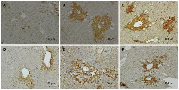
Figure 4 Immunohistological analysis of transforming growth factor-β1 (brown) in the mice livers treated with carbon tetrachloride.
A: Control; B: Carbon tetrachloride (CCl4); C: pLac + CCl4; D: pKal (100 μg) + CCl4; E: pKal (50 μg) + CCl4; F: pKal (25 μg) + CCl4.
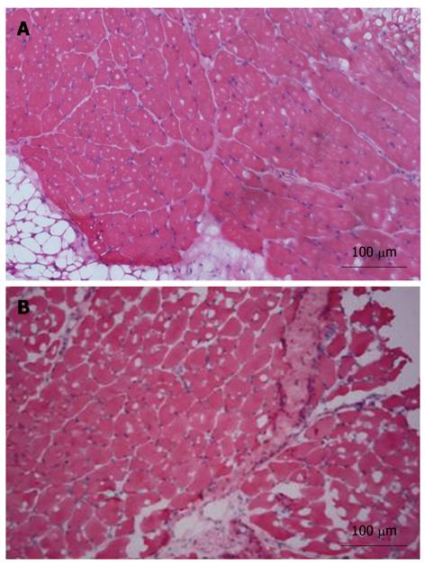
Figure 5 Inflammatory response and muscle damage arising from plasmid injection and electroporation.
A: Saline; B: pLac.
- Citation: Diao Y, Zhao XF, Lin JS, Wang QZ, Xu RA. Protection of the liver against CCl4-induced injury by intramuscular electrotransfer of a kallistatin-encoding plasmid. World J Gastroenterol 2011; 17(1): 111-117
- URL: https://www.wjgnet.com/1007-9327/full/v17/i1/111.htm
- DOI: https://dx.doi.org/10.3748/wjg.v17.i1.111


