Copyright
©2010 Baishideng.
World J Gastroenterol. Feb 28, 2010; 16(8): 973-981
Published online Feb 28, 2010. doi: 10.3748/wjg.v16.i8.973
Published online Feb 28, 2010. doi: 10.3748/wjg.v16.i8.973
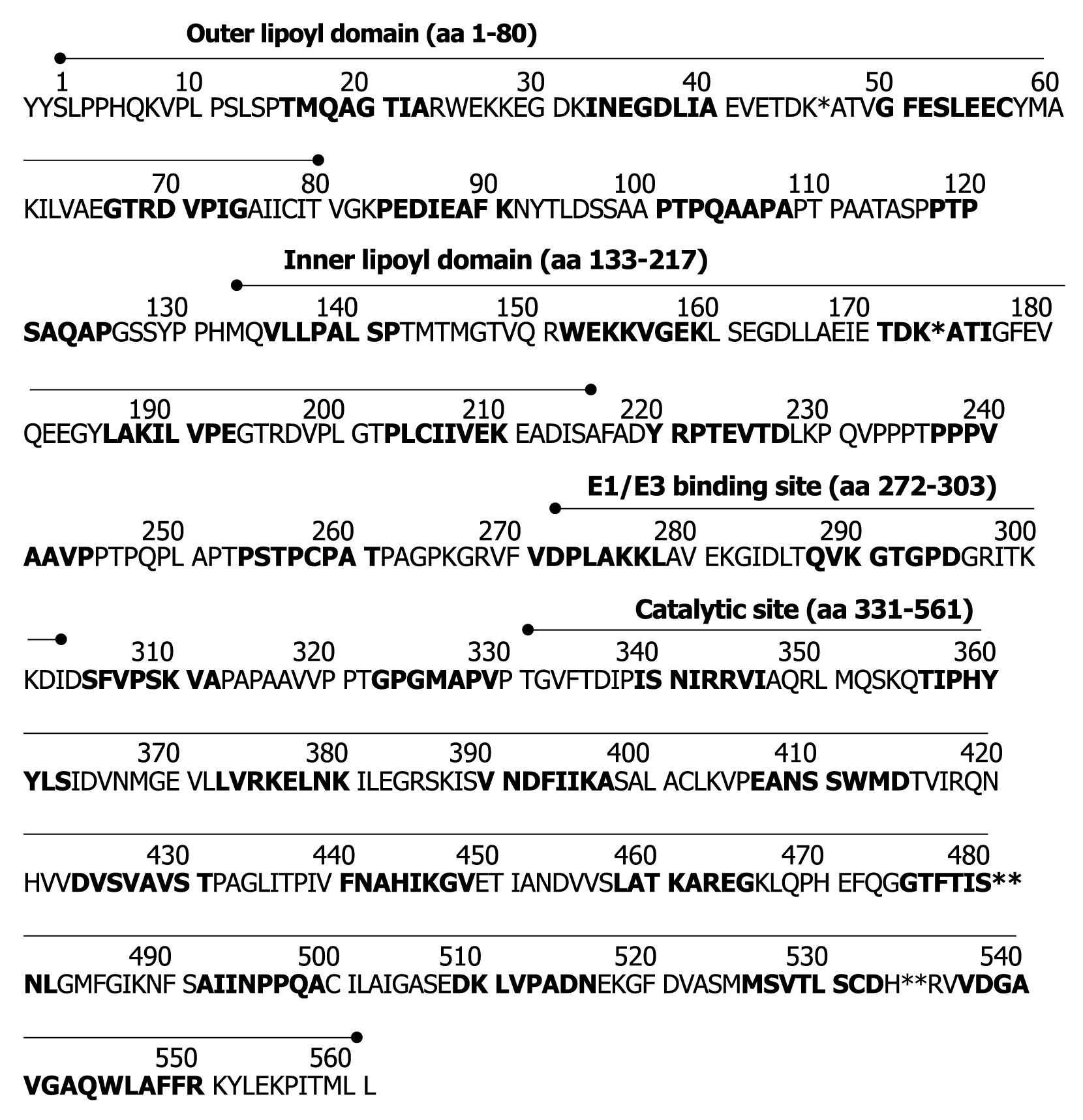
Figure 1 Amino acid (aa) sequence of the human pyruvate dehydrogenase complex E2-component (PDC-E2).
For epitope mapping 25 mer peptides with 8 overlapping amino acids were constructed (see also Table 2). Overlapping amino acids are printed in bold; amino acid sequences of the immunodominant lipoyl binding epitopes in the outer (aa 41-53) and inner lipoyl domain (aa 167-183) are underlined. *Lipoyl binding lysine in the outer and inner lipoyl domains; **Active sites within the catalytic domain (S480 and H534).
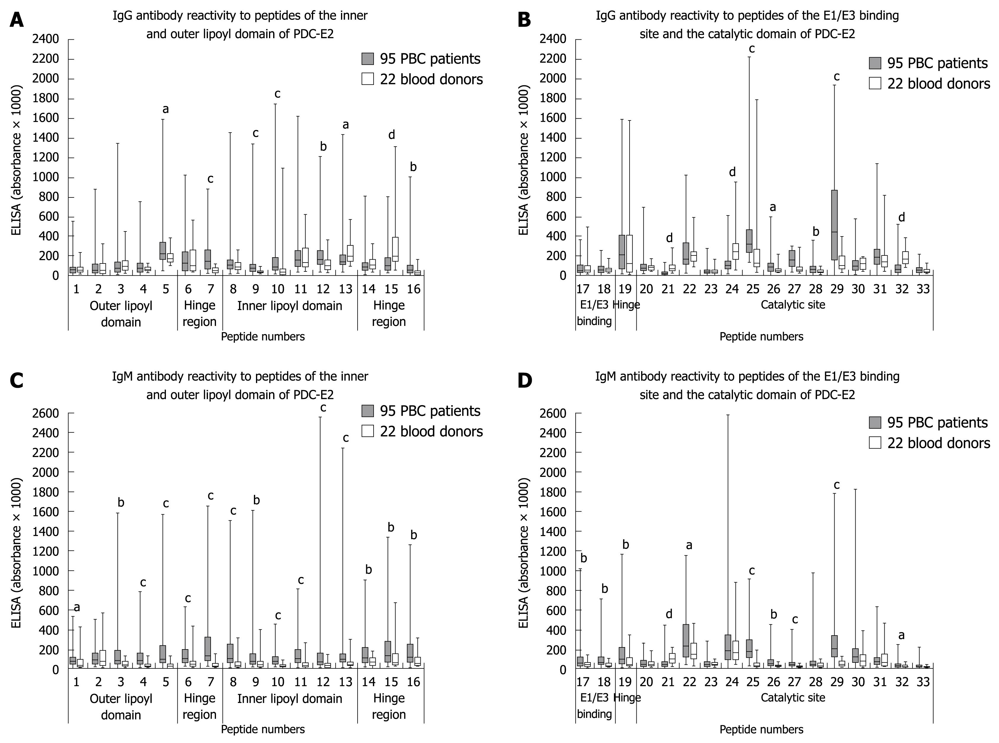
Figure 2 Box plots showing the reactivity of sera from 95 primary biliary cirrhosis (PBC) patients (grey bars) and 22 blood donors (white bars) with 33 overlapping peptides spanning the whole PDC-E2 sequence.
IgG antibody reactivities. A: Peptide 1-16; B: Peptide 17-33; IgM antibody reactivities. C: Peptide 1-16; D: Peptide 17-33. Solid bars extend from the 25th to 75th perzentile. The line in the middle is the median. The whiskers extend down to the lowest value and up to the highest value. Significantly higher antibody reactivity with PBC sera than with sera from healthy individuals: aP < 0.05, bP < 0.01, cP < 0.001. Significantly lower antibody reactivity with PBC sera than with sera from healthy individuals: dP < 0.001. ELISA: Enzyme-linked immunosorbent assay.
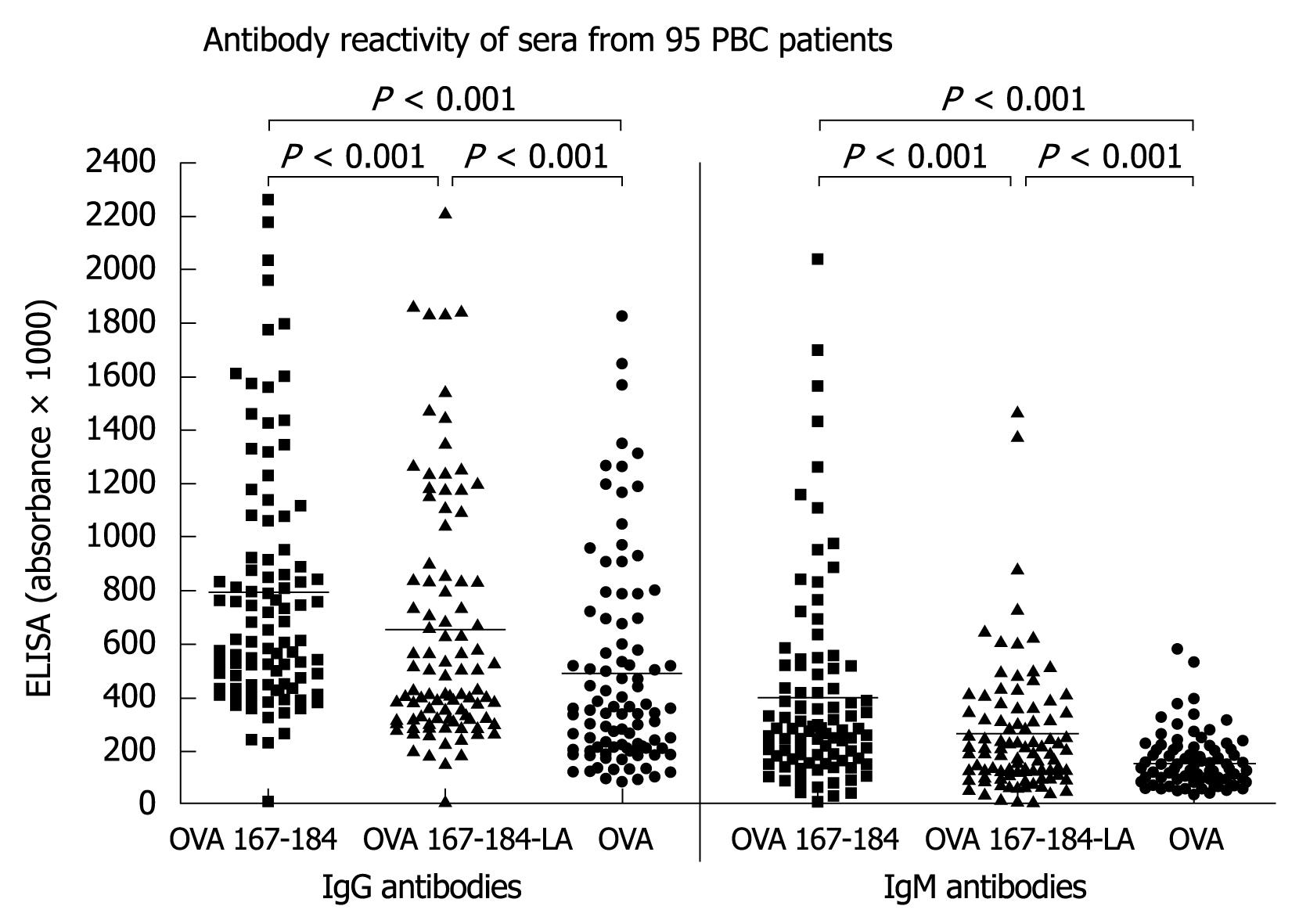
Figure 3 IgG- and IgM-reactivity of sera from 95 PBC patients with the OVA coupled unlipoylated and lipoylated peptide 167-184 (OVA-167-184 and OVC-167-184-LA) (without subtraction of the OVA-values) and OVA alone.
- Citation: Braun S, Berg C, Buck S, Gregor M, Klein R. Catalytic domain of PDC-E2 contains epitopes recognized by antimitochondrial antibodies in primary biliary cirrhosis. World J Gastroenterol 2010; 16(8): 973-981
- URL: https://www.wjgnet.com/1007-9327/full/v16/i8/973.htm
- DOI: https://dx.doi.org/10.3748/wjg.v16.i8.973


