Copyright
©2010 Baishideng.
World J Gastroenterol. Feb 28, 2010; 16(8): 1008-1013
Published online Feb 28, 2010. doi: 10.3748/wjg.v16.i8.1008
Published online Feb 28, 2010. doi: 10.3748/wjg.v16.i8.1008
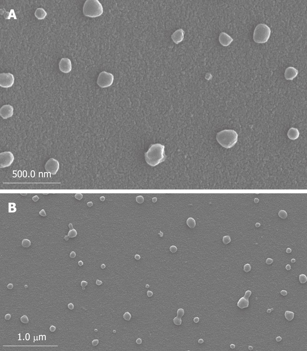
Figure 1 Scanning electronic microphotographs of poly (ethylene glycol)-block-poly (D,L-lactide) (PEG-PDLLA) nanovesicles.
A: × 40 000; B: × 20 000.
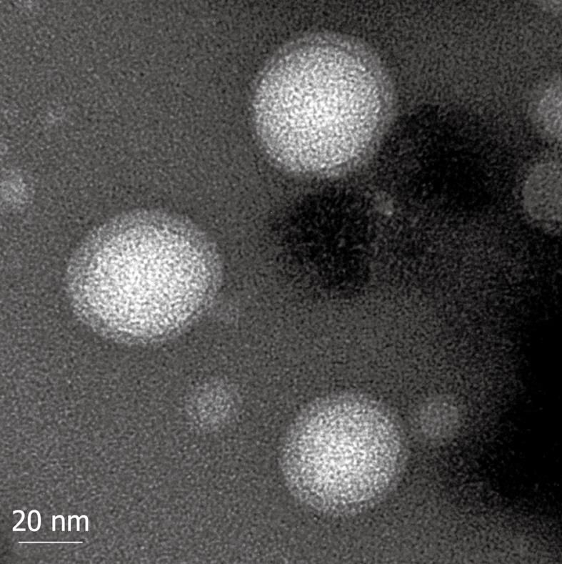
Figure 2 Transmission electron micrographs of PEG-PDLLA nanovesicles.
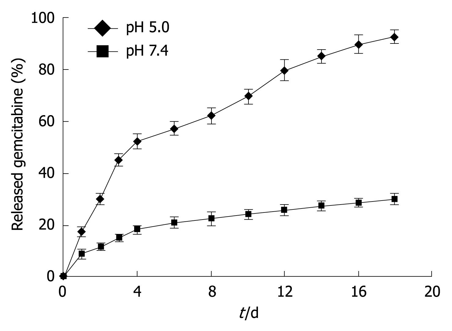
Figure 3 Release of gemcitabine from nanovesicles at pH 7.
4 and 5.0. Data are presented as mean ± SD. P < 0.05 at every point of pH 5.0 vs pH 7.4.
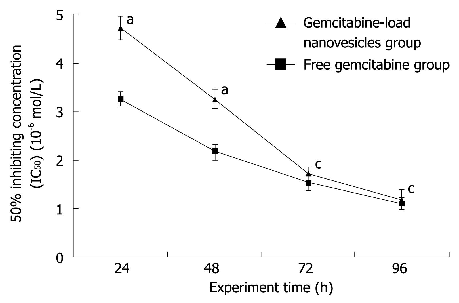
Figure 4 IC50 of gemcitabine-loaded nanovesicles and free gemcitabine in human SW1990 pancreatic cancer cells.
Data are presented as mean ± SD. aP < 0.05, cP > 0.05 vs free gemcitabine group.
-
Citation: Jia L, Zheng JJ, Jiang SM, Huang KH. Preparation, physicochemical characterization and cytotoxicity
in vitro of gemcitabine-loaded PEG-PDLLA nanovesicles. World J Gastroenterol 2010; 16(8): 1008-1013 - URL: https://www.wjgnet.com/1007-9327/full/v16/i8/1008.htm
- DOI: https://dx.doi.org/10.3748/wjg.v16.i8.1008


