Copyright
©2010 Baishideng.
World J Gastroenterol. Feb 14, 2010; 16(6): 778-781
Published online Feb 14, 2010. doi: 10.3748/wjg.v16.i6.778
Published online Feb 14, 2010. doi: 10.3748/wjg.v16.i6.778
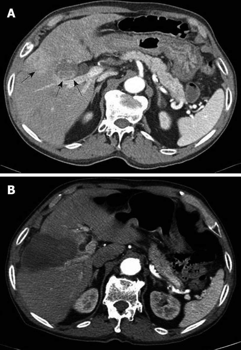
Figure 1 Computed tomography (CT) scan of the patient.
A: CT scan in an arterial phase demonstrates a 3.5 cm hepatocellular carcinoma lesion (arrows) at segment IV of the liver adjacent to the gallbladder, posterior to the lesion where a previous percutaneous ethanol injection has been performed; B: CT scan obtained after radiofrequency thermal ablation reveals complete ablation for the hepatocellular carcinoma without apparent complications such as a gallbladder injury on the immediate follow-up.
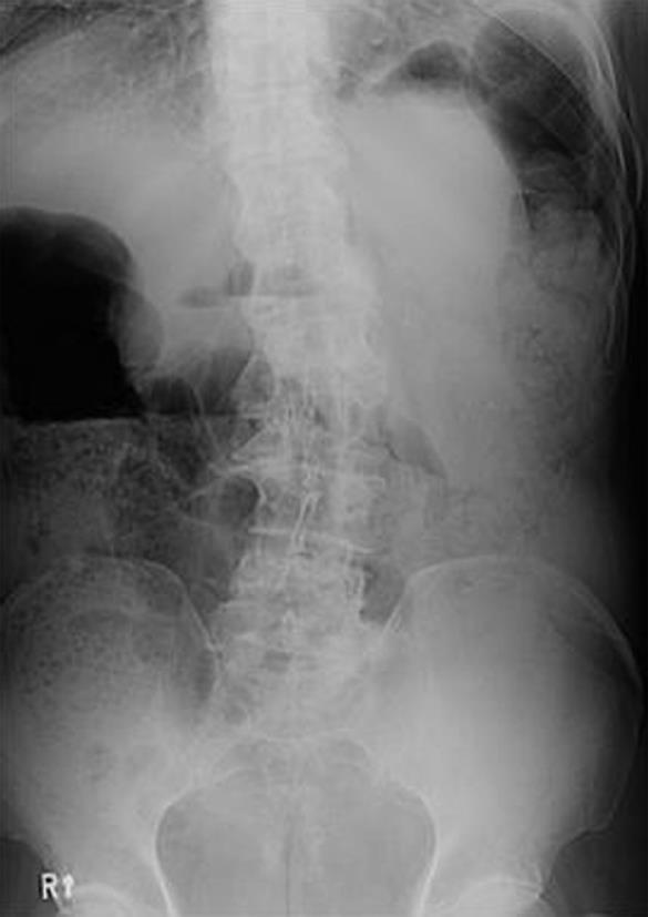
Figure 2 A simple abdominal radiograph reveals no evidence of bowel perforation, except a distension of the large intestine.
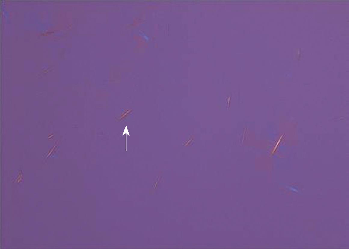
Figure 3 Strongly negative birefringent, needle-shaped monosodium urate crystals (arrow) in synovial fluid from a patient under compensated polarized light.
- Citation: Choi DH, Lee HS. A case of gouty arthritis following percutaneous radiofrequency ablation for hepatocellular carcinoma. World J Gastroenterol 2010; 16(6): 778-781
- URL: https://www.wjgnet.com/1007-9327/full/v16/i6/778.htm
- DOI: https://dx.doi.org/10.3748/wjg.v16.i6.778


