Copyright
©2010 Baishideng.
World J Gastroenterol. Feb 14, 2010; 16(6): 683-690
Published online Feb 14, 2010. doi: 10.3748/wjg.v16.i6.683
Published online Feb 14, 2010. doi: 10.3748/wjg.v16.i6.683
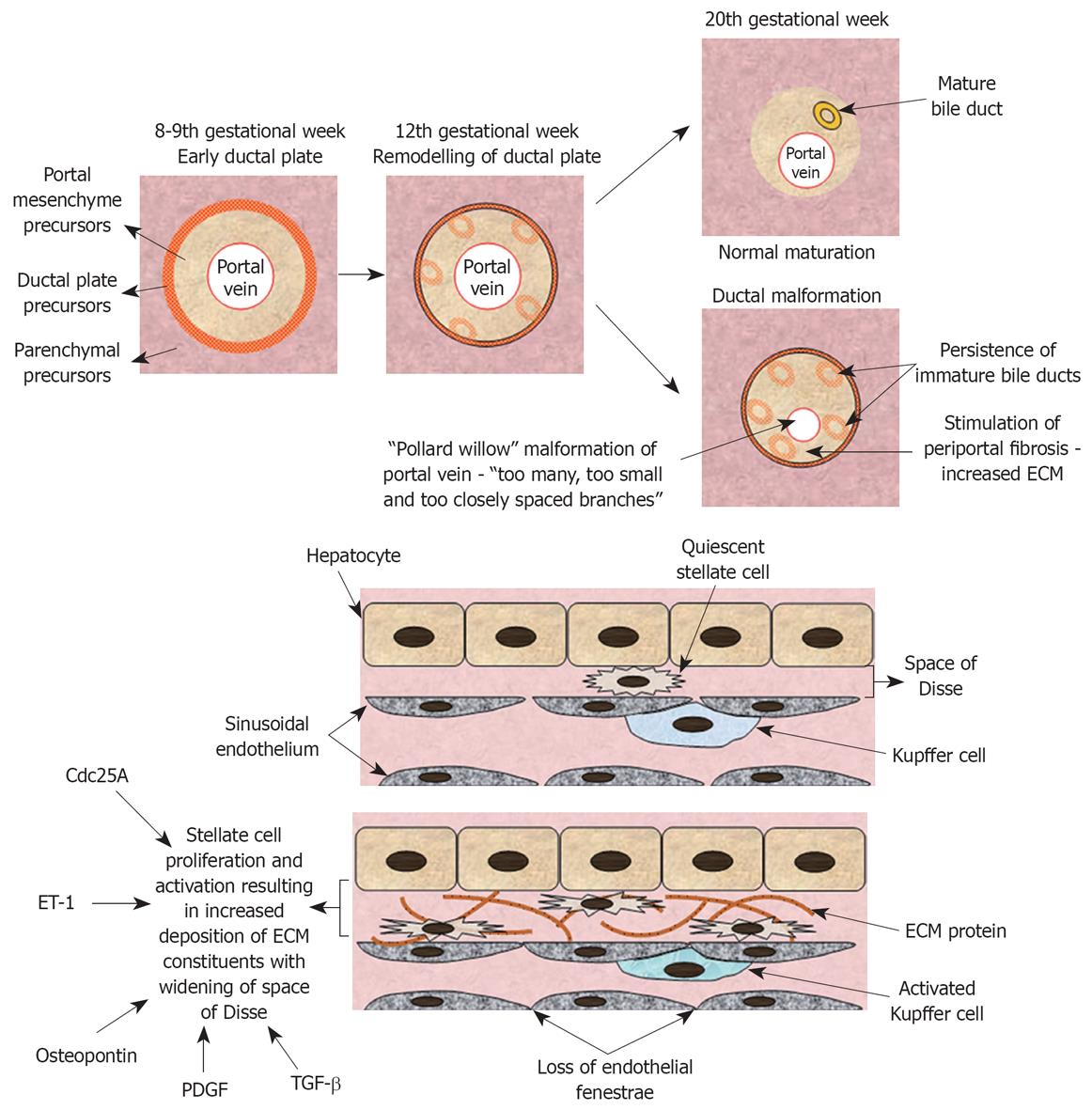
Figure 1 Pathogenesis of congenital hepatic fibrosis.
Embryological and molecular perspective. ET-1: Endothelin 1;
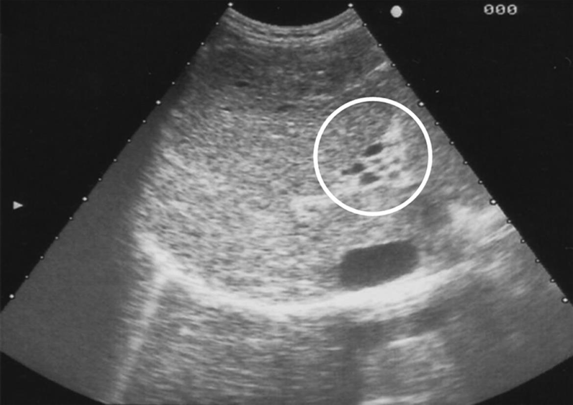
Figure 2 Ultrasound image of a patient with congenital hepatic fibrosis (CHF).
Heterogenous appearance of hepatic parenchyma. The circled area depicts the presence of portal vein cavernous transformation.
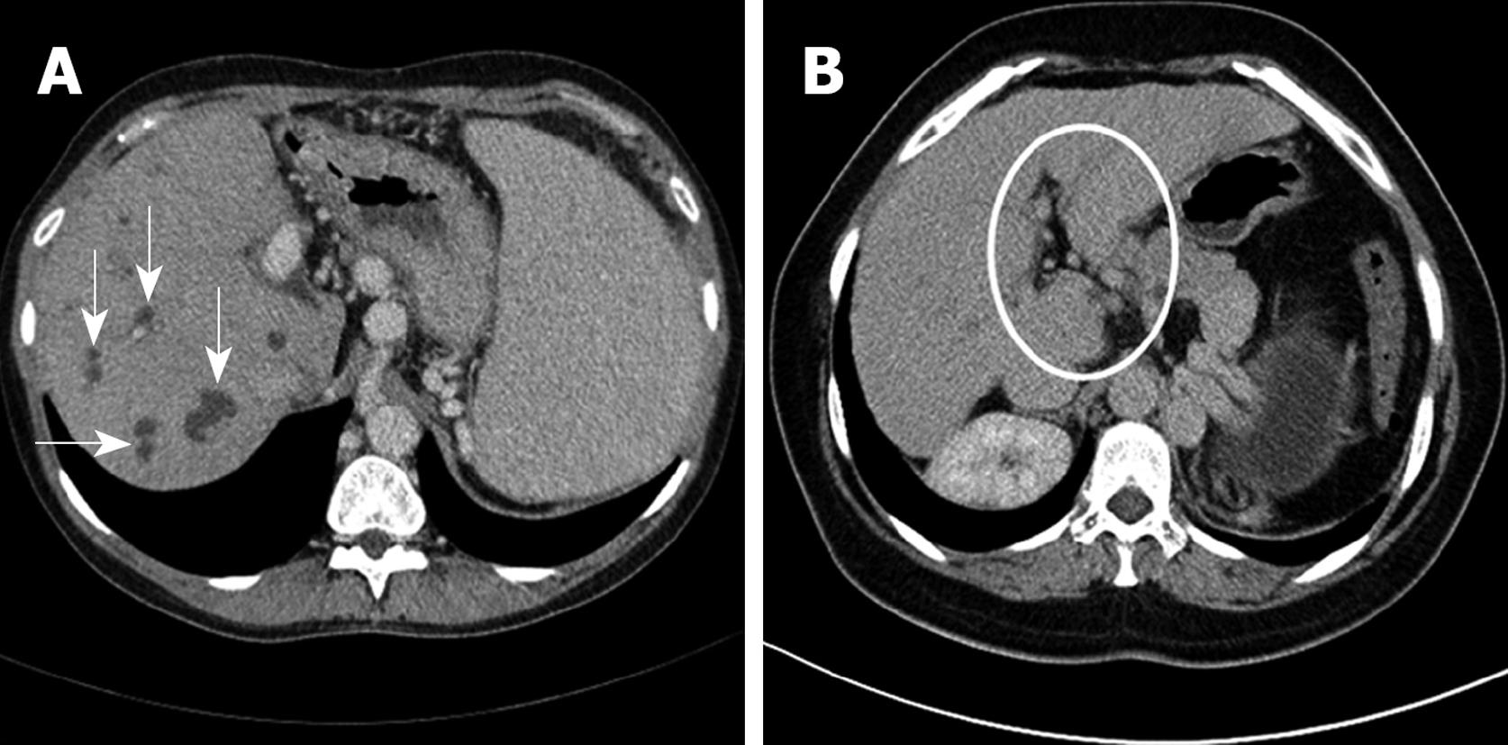
Figure 3 Abdominal computerized tomography (CT) scans of two patients with CHF.
A: White arrows depict cystic dilatations of the biliary tree associated with Caroli's syndrome; B: Circled area shows portal vein cavernous transformation in a patient with Bardet-Biedl syndrome.
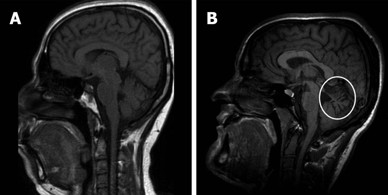
Figure 4 Brain magnetic resonance imaging (MRI) scans of two patients with congenital hepatic fibrosis.
A: A patient with Bardet-Biedl syndrome with normal findings; B: The circled area depicts cerebellar vermis atrophy manifested by more prominent folds/sulci, associated with Joubert syndrome.
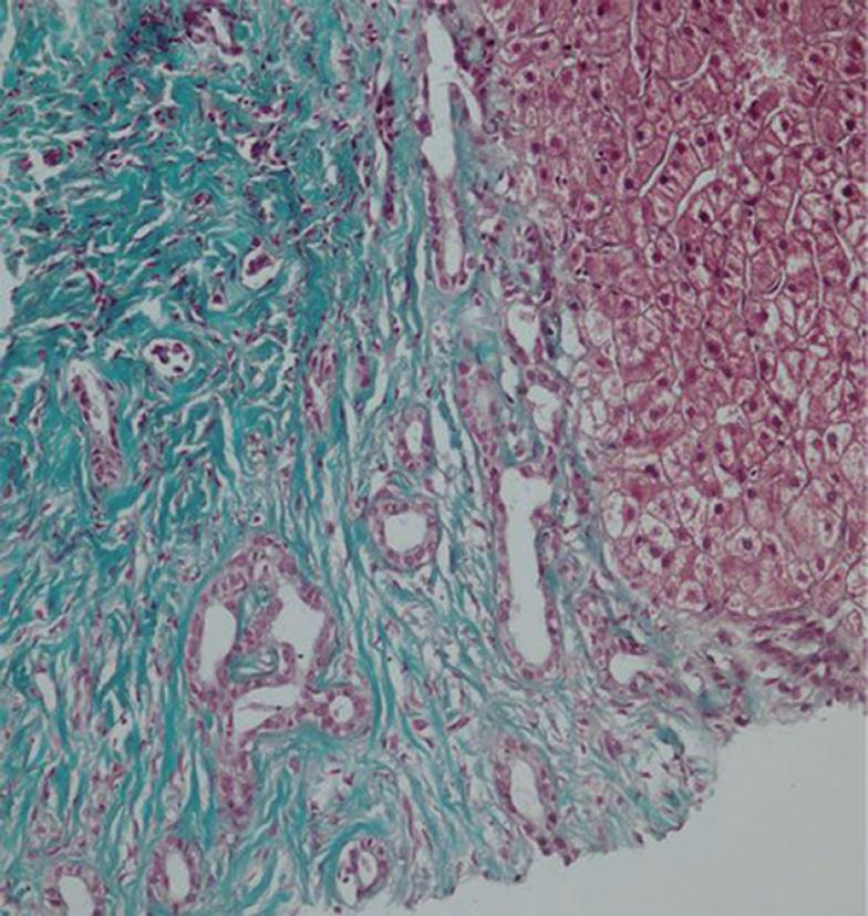
Figure 5 Liver biopsy of a patient with CHF.
The left side of the image depicts a portal area with extensive fibrosis and the presence of several bile ducts with cuboidal epithelium that have arrested at different stages of the maturation process. On the right, hepatocytes with normal morphology may be seen (× 230, trichrome stain).
- Citation: Shorbagi A, Bayraktar Y. Experience of a single center with congenital hepatic fibrosis: A review of the literature. World J Gastroenterol 2010; 16(6): 683-690
- URL: https://www.wjgnet.com/1007-9327/full/v16/i6/683.htm
- DOI: https://dx.doi.org/10.3748/wjg.v16.i6.683


