Copyright
©2010 Baishideng.
World J Gastroenterol. Feb 7, 2010; 16(5): 648-651
Published online Feb 7, 2010. doi: 10.3748/wjg.v16.i5.648
Published online Feb 7, 2010. doi: 10.3748/wjg.v16.i5.648
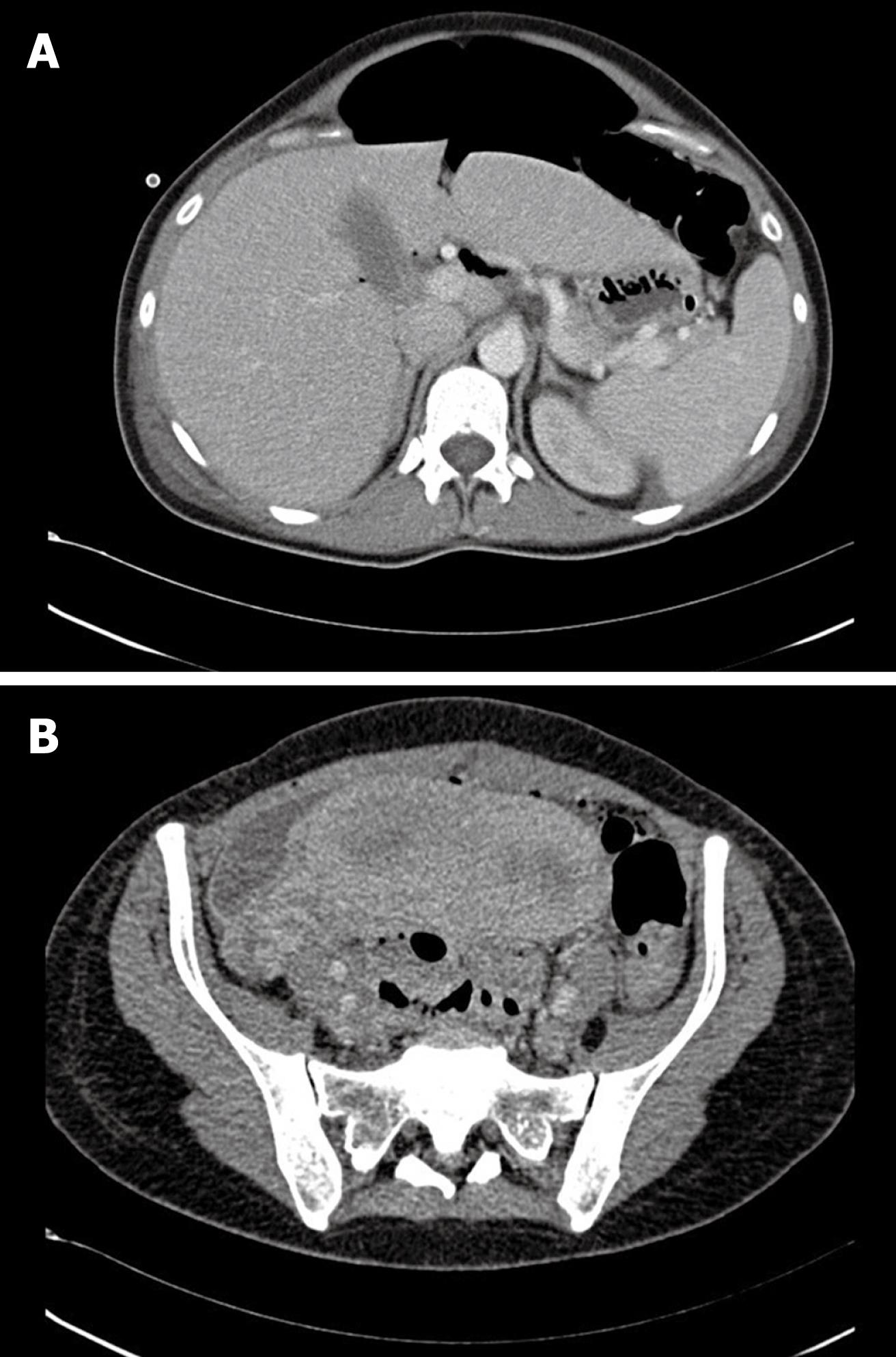
Figure 1 Axial computed tomography (CT) scan.
A: Free air in the abdominal cavity; B: Pelvic abscess at the right side of the enlarged uterus.
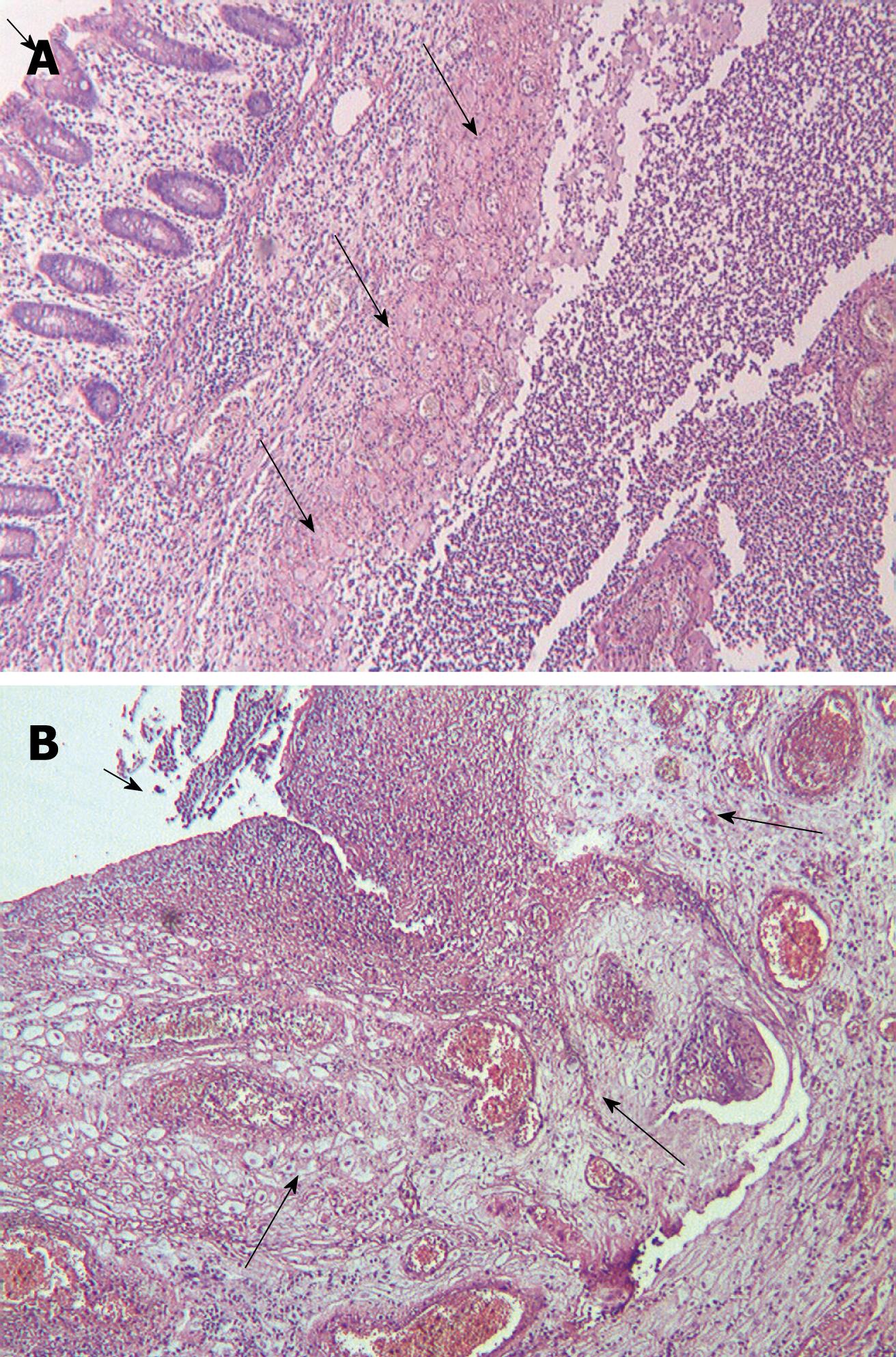
Figure 2 The rectal wall.
A: Decidualization of the rectal wall (long arrows); mucosa side of the rectal wall (short arrow) (HE, × 40); B: Decidualized endometriosis around the rectal perforation (long arrows); rectal perforation with necrosis at the peritoneal side of the rectal wall (short arrow) (HE, × 100).
- Citation: Pisanu A, Deplano D, Angioni S, Ambu R, Uccheddu A. Rectal perforation from endometriosis in pregnancy: Case report and literature review. World J Gastroenterol 2010; 16(5): 648-651
- URL: https://www.wjgnet.com/1007-9327/full/v16/i5/648.htm
- DOI: https://dx.doi.org/10.3748/wjg.v16.i5.648


