Copyright
©2010 Baishideng.
World J Gastroenterol. Feb 7, 2010; 16(5): 636-640
Published online Feb 7, 2010. doi: 10.3748/wjg.v16.i5.636
Published online Feb 7, 2010. doi: 10.3748/wjg.v16.i5.636
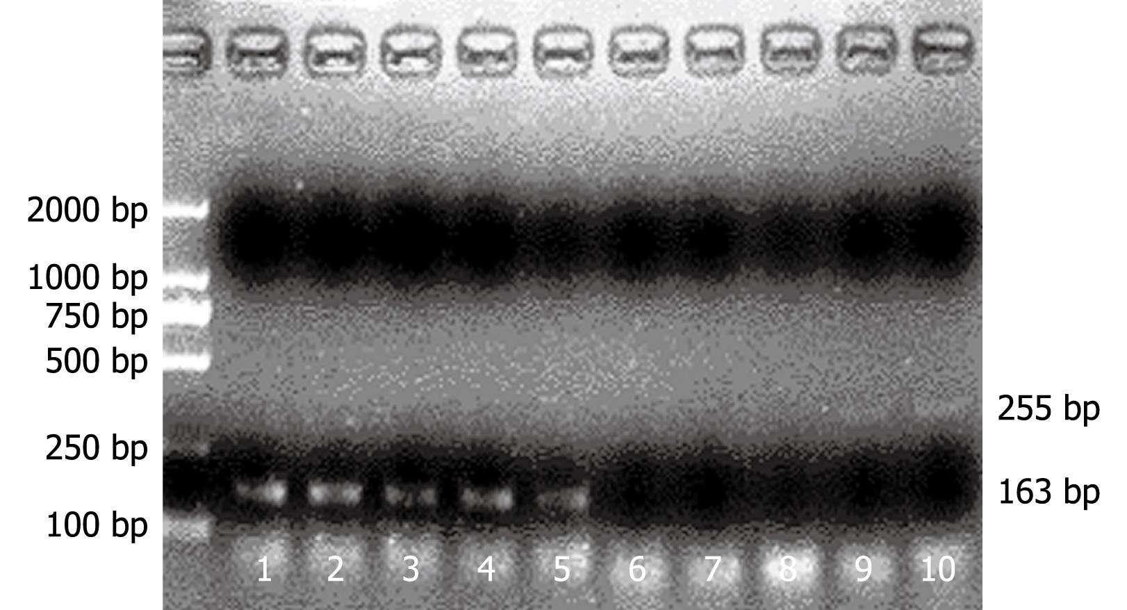
Figure 1 Reverse transcription polymerase chain reaction (RT-PCR) shows indoleamine 2,3-dioxygenase (IDO) and FasL expression in Kupffer cells (KC) before and after activation with recombinant interferon-γ (IFN-γ).
RT-PCR showing FasL expression in KC before (lanes 1 and 2) and after activation for 6 h (lanes 3 and 4) or 24 h (lane 5) with IFN-γ. RT-PCR showing IDO expression in KC before (lanes 6-8) and after activation for 6 h (lane 9) or 24 h (lane 10) with IFN-γ.
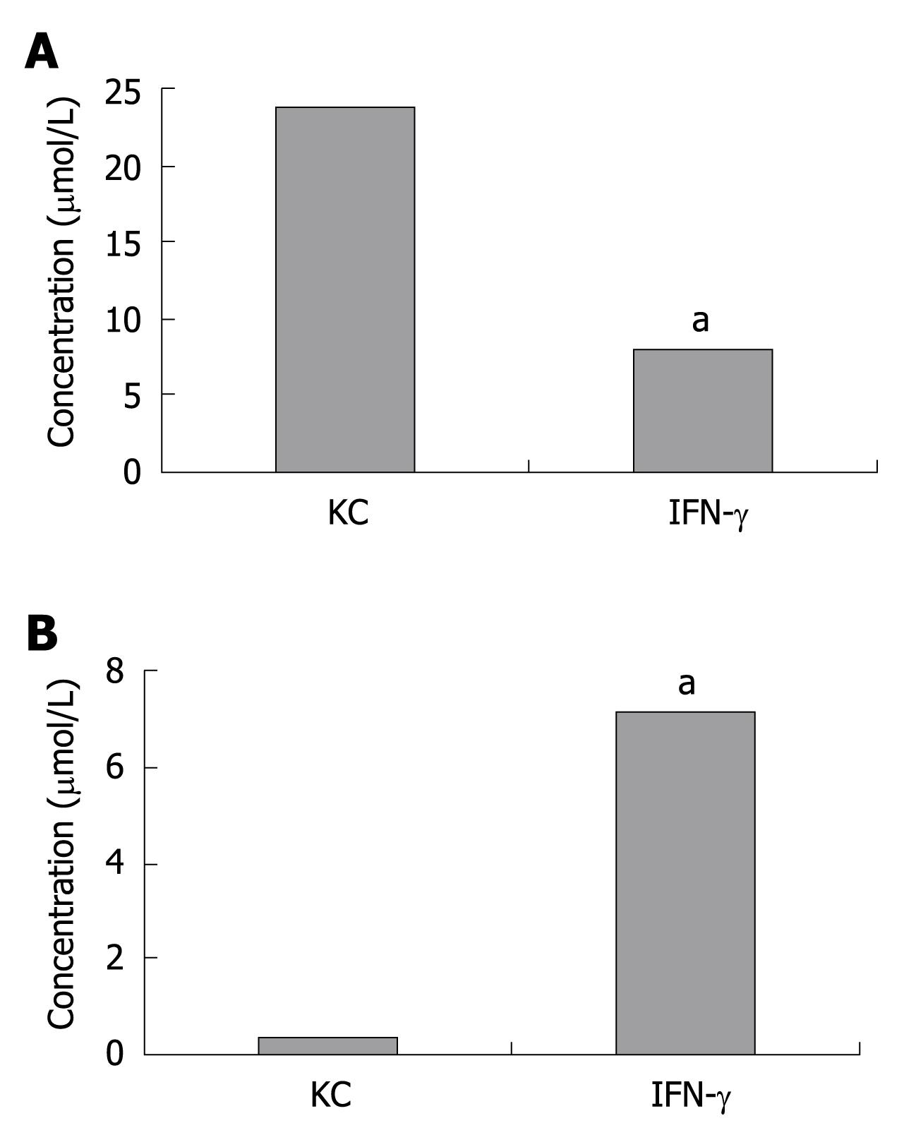
Figure 2 Tryptophan (A) and kynurenine (B) concentration in cultures of KC.
The kynurenine concentration significantly increased while tryptophan decreased in KC pretreated with IFN-γ for 24 h. aP < 0.05 vs KC group.
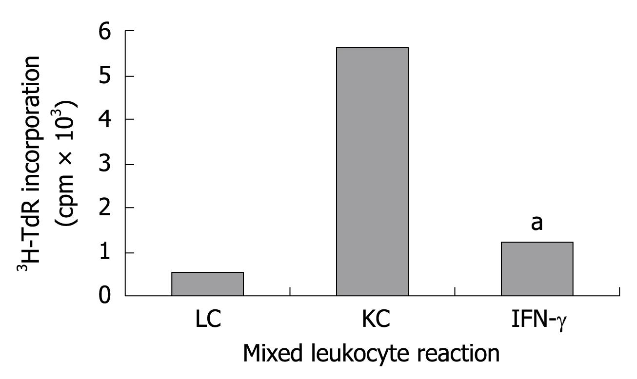
Figure 3 Reduction of allogeneic T-cell stimulation by IDO and FasL-expressing KC.
Naive lymphocyte (LC), naive KC, KC pretreated with IFN-γ were coincubated with allogeneic spleen T-cell for 5 d and cell proliferation measured by 3H-TdR incorporation. aP < 0.05 vs KC group.
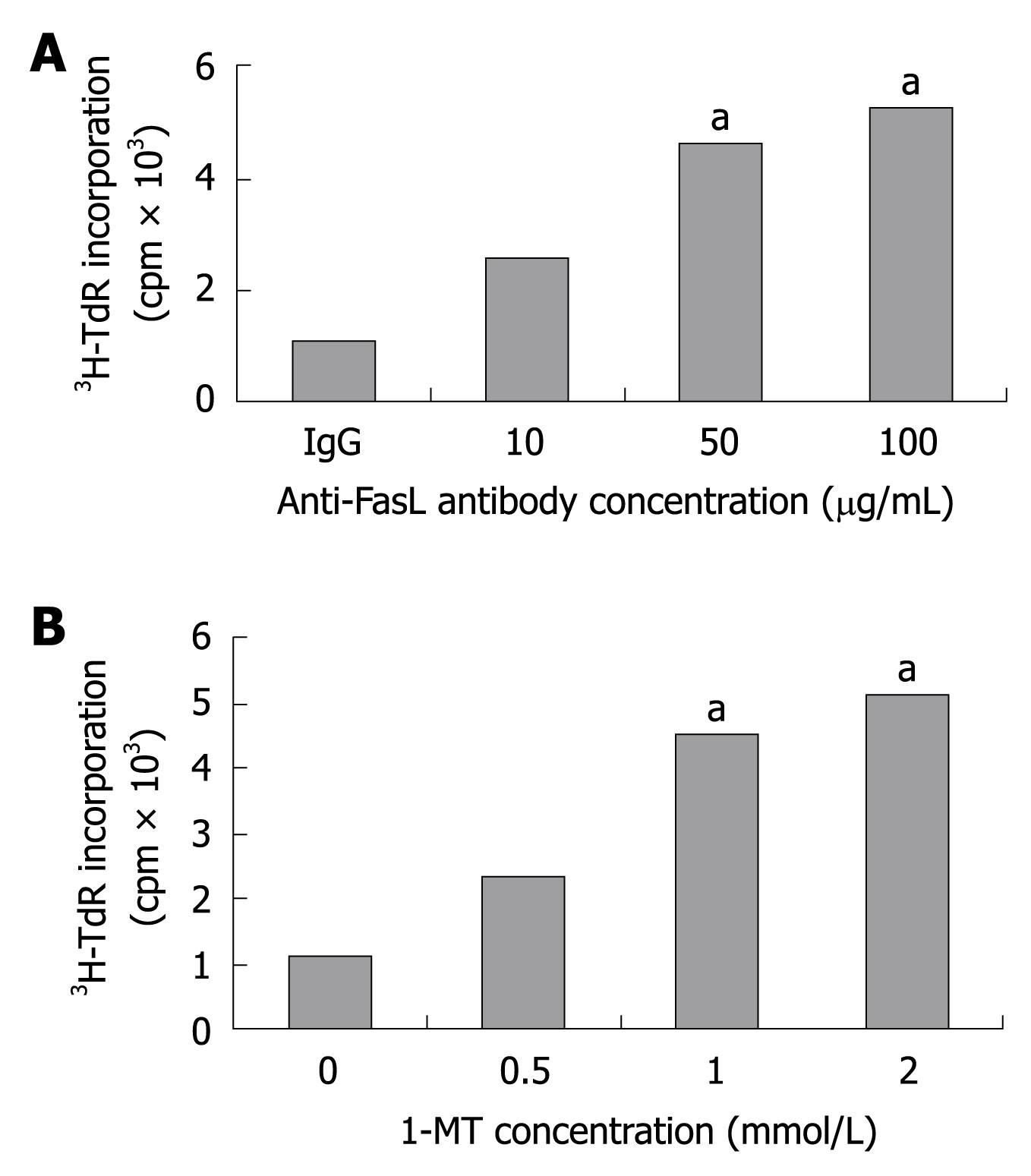
Figure 4 Anti-FasL antibody (A) or 1-methyl-tryptophan (1-MT) (B) enhances the proliferation of allogeneic T-cell stimulated with KC pretreated with IFN-γ.
aP < 0.05 vs control group.
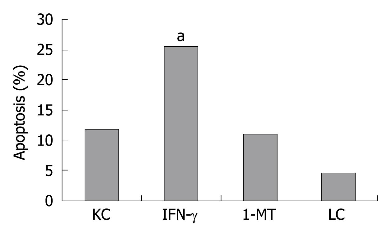
Figure 5 KC induces apoptosis of allogeneic T-cell.
The KC pretreated with IFN-γ exhibit a significant allogeneic T-cell apoptosis. aP < 0.05 vs KC group.
- Citation: Yan ML, Wang YD, Tian YF, Lai ZD, Yan LN. Inhibition of allogeneic T-cell response by Kupffer cells expressing indoleamine 2,3-dioxygenase. World J Gastroenterol 2010; 16(5): 636-640
- URL: https://www.wjgnet.com/1007-9327/full/v16/i5/636.htm
- DOI: https://dx.doi.org/10.3748/wjg.v16.i5.636


