Copyright
©2010 Baishideng.
World J Gastroenterol. Feb 7, 2010; 16(5): 538-546
Published online Feb 7, 2010. doi: 10.3748/wjg.v16.i5.538
Published online Feb 7, 2010. doi: 10.3748/wjg.v16.i5.538
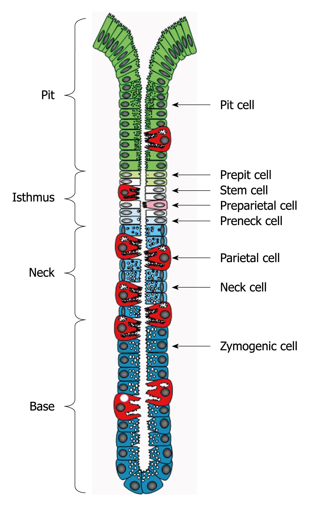
Figure 1 Diagrammatic representation of the gastric unit or gland.
It is composed of 4 regions: pit, isthmus, neck and base and populated by a heterogeneous population of cell types. Pit, parietal and neck cells originate in the isthmus from stem (granule-free) cells and their immediate descendants (prepit, preparietal and preneck cells). Neck cells are not end cells and therefore, at the neck-base border, they transform into zymogenic cells.
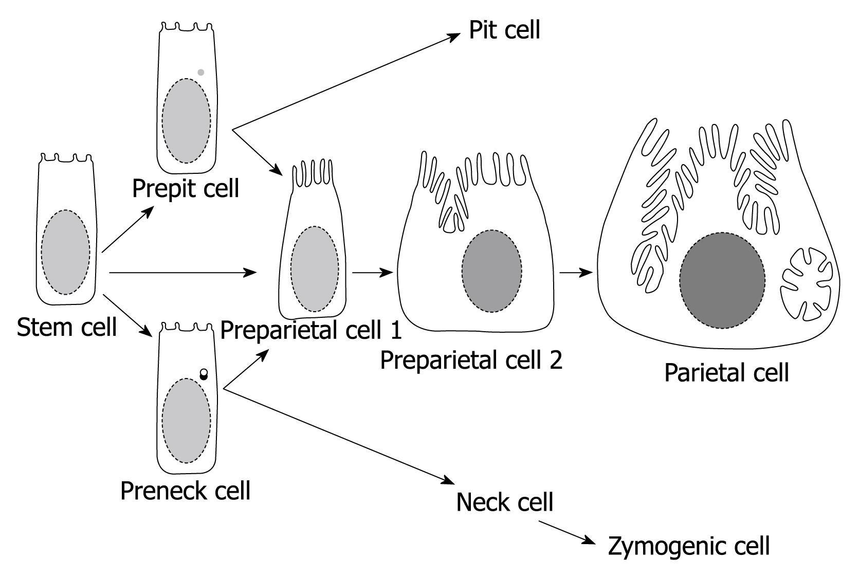
Figure 2 Diagram summarizing the origin and differentiation program of parietal cells.
The stem cell either directly or indirectly (via prepit or preneck cells) give rise to the committed progenitors of parietal cells (preparietal cells). Preparietal cells evolve in 4 successive stages (2 are depicted here) to become mature parietal cells. Preparietal cell 1 is characterized by long microvilli and preparietal cell 2 by an incipient canaliculus which expands around the nucleus in the mature parietal cell.
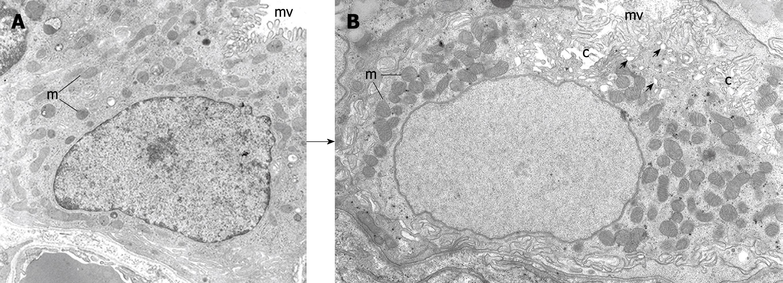
Figure 3 Electron micrographs depicting 2 stages of the differentiating preparietal cells as seen in the human stomach.
Original magnification, × 12 000. A: Preparietal cell showing relatively long numerous microvilli (mv). The mitochondria (m) are relatively few and small; B: Preparietal cell showing an incipient canaliculus (c) and relatively numerous mitochondria (m) which appear larger than those in A. The apical cytoplasm shows few tubulovesicles (small arrows) and long numerous microvilli (mv). A is reproduced from Karam et al[4] with permission from Wiley-Blackwell/AlphaMed press.
- Citation: Karam SM. A focus on parietal cells as a renewing cell population. World J Gastroenterol 2010; 16(5): 538-546
- URL: https://www.wjgnet.com/1007-9327/full/v16/i5/538.htm
- DOI: https://dx.doi.org/10.3748/wjg.v16.i5.538


