Copyright
©2010 Baishideng Publishing Group Co.
World J Gastroenterol. Dec 7, 2010; 16(45): 5722-5726
Published online Dec 7, 2010. doi: 10.3748/wjg.v16.i45.5722
Published online Dec 7, 2010. doi: 10.3748/wjg.v16.i45.5722
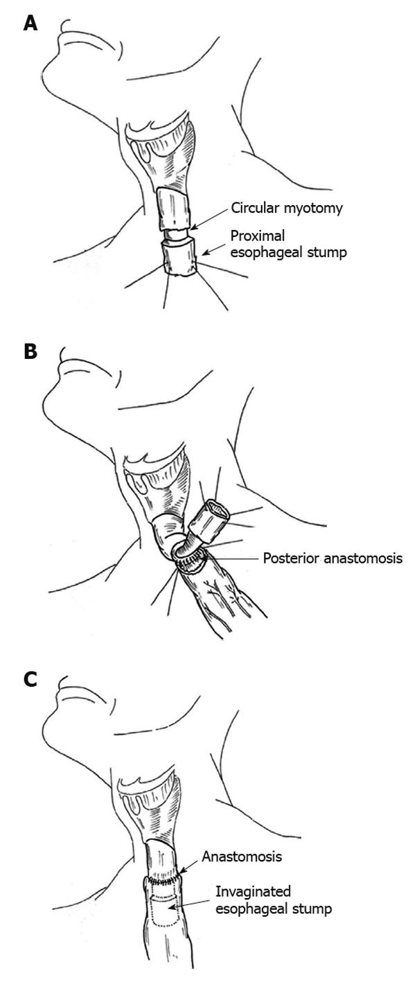
Figure 1 Esophagogastric anastomosis with stomach invagination.
A: Diagram showing the circular myotomy (long arrow) in the section of the proximal esophageal stump (short arrow), which created a 4.0 cm segment of extension to be invaginated into the stomach (the illustration of the trachea was omitted); B: Diagram showing the anastomosis of the posterior wall of the esophagus performed first using interrupted sutures (the illustration of the trachea was omitted); C: Diagram showing the sectioned esophagus protruding into the stomach (the illustration of the trachea was omitted).
- Citation: Henriques AC, Godinho CA, Saad Jr R, Waisberg DR, Zanon AB, Speranzini MB, Waisberg J. Esophagogastric anastomosis with invagination into stomach: New technique to reduce fistula formation. World J Gastroenterol 2010; 16(45): 5722-5726
- URL: https://www.wjgnet.com/1007-9327/full/v16/i45/5722.htm
- DOI: https://dx.doi.org/10.3748/wjg.v16.i45.5722


