Copyright
©2010 Baishideng Publishing Group Co.
World J Gastroenterol. Nov 28, 2010; 16(44): 5611-5615
Published online Nov 28, 2010. doi: 10.3748/wjg.v16.i44.5611
Published online Nov 28, 2010. doi: 10.3748/wjg.v16.i44.5611
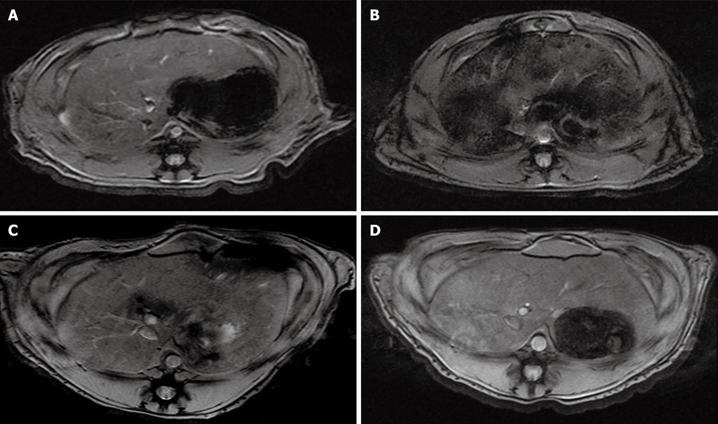
Figure 1 T2*-weighted gradient-echo magnetic resonance images show signal intensity changes of the liver after injecting superparamagnetic iron oxide-labeled human mesenchymal stem cells immediately before (A), 2 h (B) and 1 d (C), 7 d (D) after injection.
Changes in signal intensity in the liver normalized to that of back muscle at 2 h, 1 d, and 7 d were 41.87% ± 9.63%, 10.42% ± 4.3%, and 3.75% ± 1.2%, respectively (P < 0.001). Note a significant signal drop of the liver at 2 h after injecting superparamagnetic iron oxide-labeled mesenchymal stem cells and gradual return of signal intensity compared with signal intensity before transplantation.
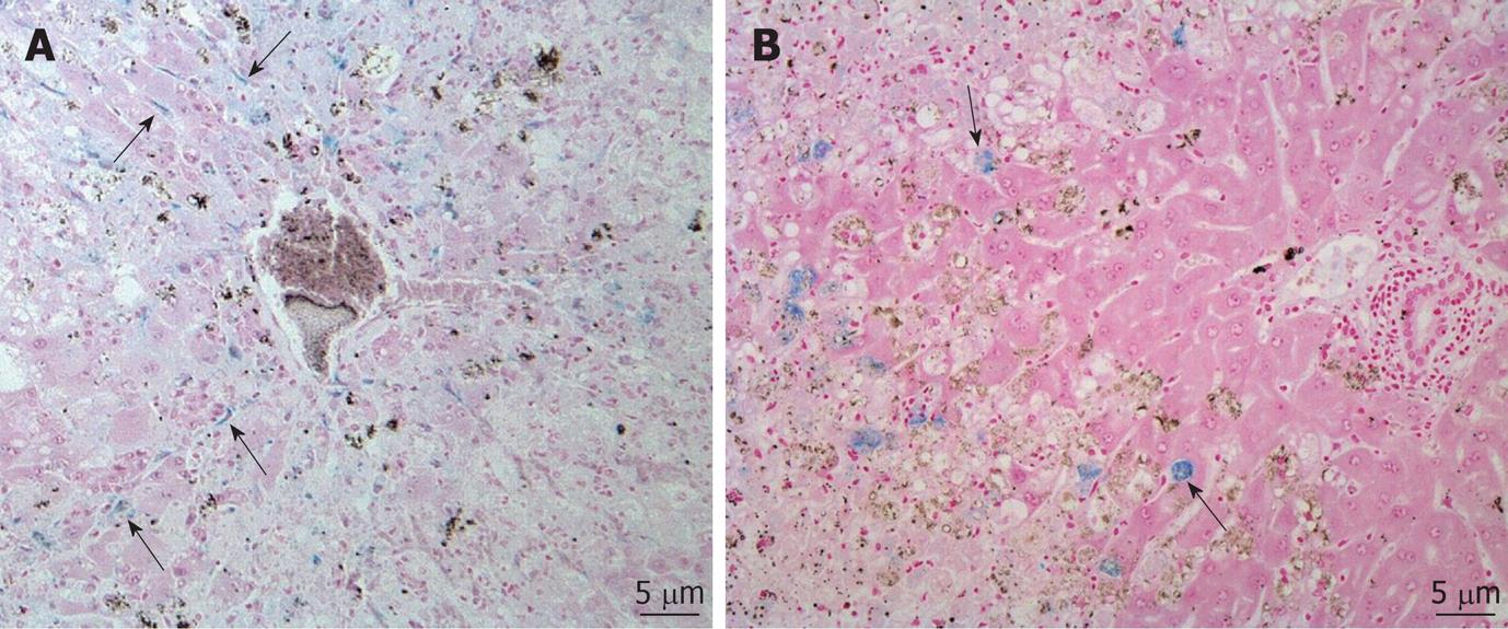
Figure 2 Photomicrographs of rabbit liver obtained after superparamagnetic iron oxide-labeled human mesenchymal stem cells injection via the mesenteric vein.
A: Section obtained on day 1 shows distributions of Prussian blue stain-positive mesenchymal stem cells (MSCs) (arrows) in the liver parenchyma along the sinusoids; B: Section obtained on day 7 shows the localization of superparamagnetic iron oxide-labeled MSCs (arrows) in the border zone between normal liver parenchyma and hepatic injury areas.
- Citation: Son KR, Chung SY, Kim HC, Kim HS, Choi SH, Lee JM, Moon WK. MRI of magnetically labeled mesenchymal stem cells in hepatic failure model. World J Gastroenterol 2010; 16(44): 5611-5615
- URL: https://www.wjgnet.com/1007-9327/full/v16/i44/5611.htm
- DOI: https://dx.doi.org/10.3748/wjg.v16.i44.5611


