Copyright
©2010 Baishideng Publishing Group Co.
World J Gastroenterol. Nov 28, 2010; 16(44): 5592-5597
Published online Nov 28, 2010. doi: 10.3748/wjg.v16.i44.5592
Published online Nov 28, 2010. doi: 10.3748/wjg.v16.i44.5592
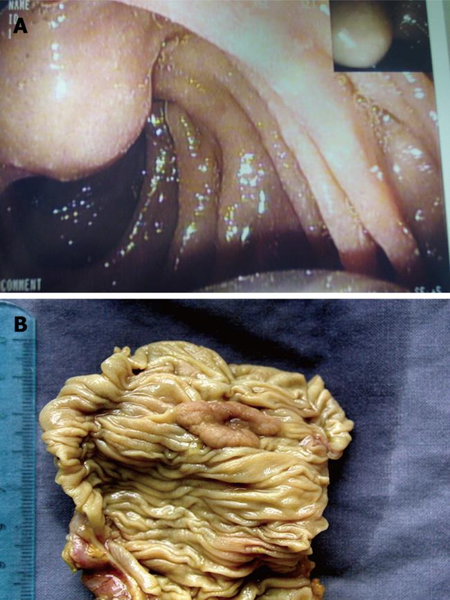
Figure 1 Endoscopic aspect (A) and resected surgical specimen (B) of a small ampullary tumor.
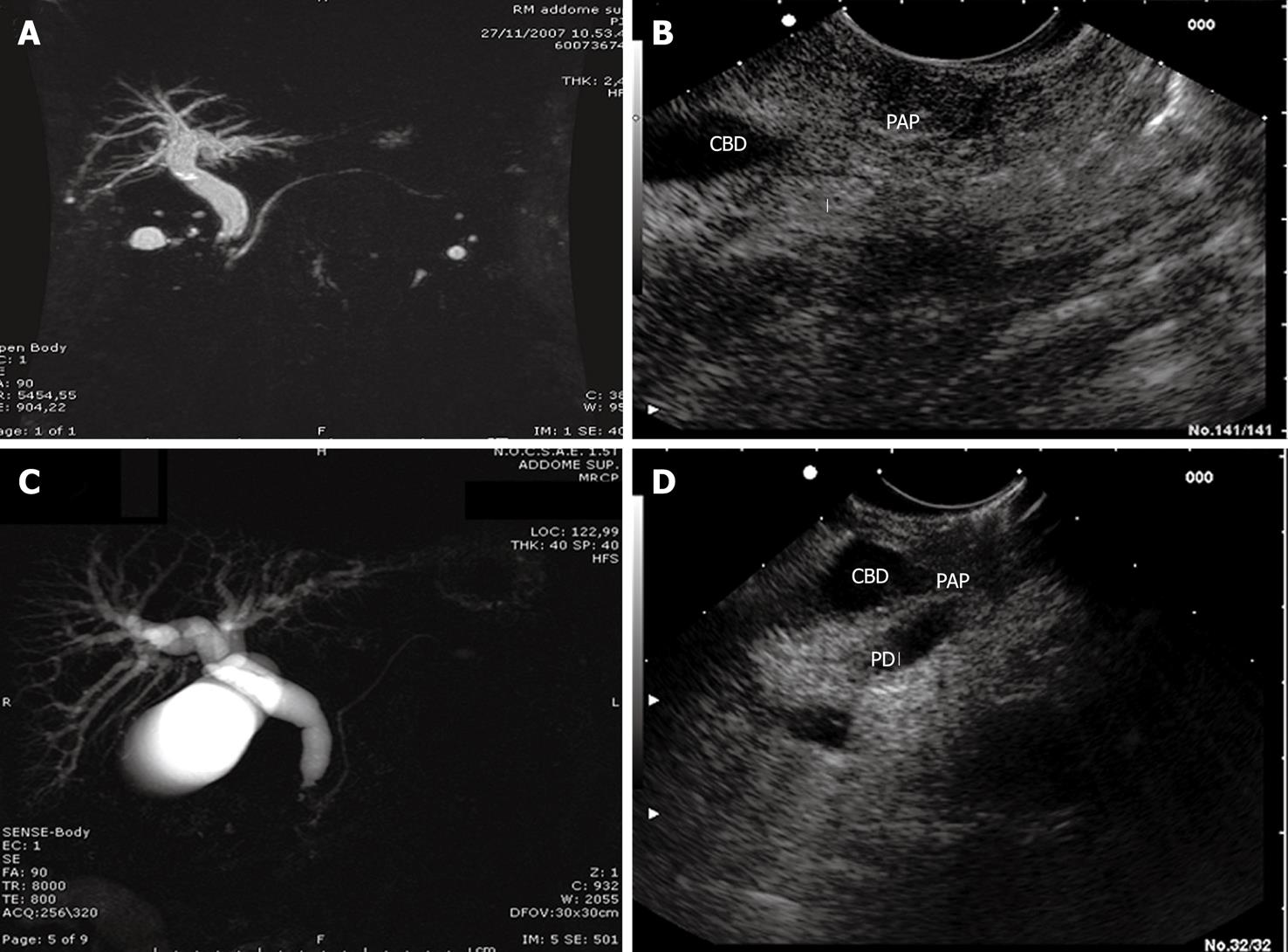
Figure 2 Ampullary tumor, stage 1.
A: Magnetic resonance imaging (MRI) showed dilation of the common bile duct and Wirsung’s duct, without space-occupying lesions of the ampulla; B: Linear endoscopic ultrasound (L-EUS) of the same patient. Ampullary tumor, stage 2; C: MRI showed dilation of the common bile duct and normal appearance of Wirsung’s duct; D: L-EUS scan of the same patient, which showed duodenal wall disruption without pancreas invasion. CBD: Common bile duct; PAP: Papilla (Vater's papilla); PD: Pancreatic duct.
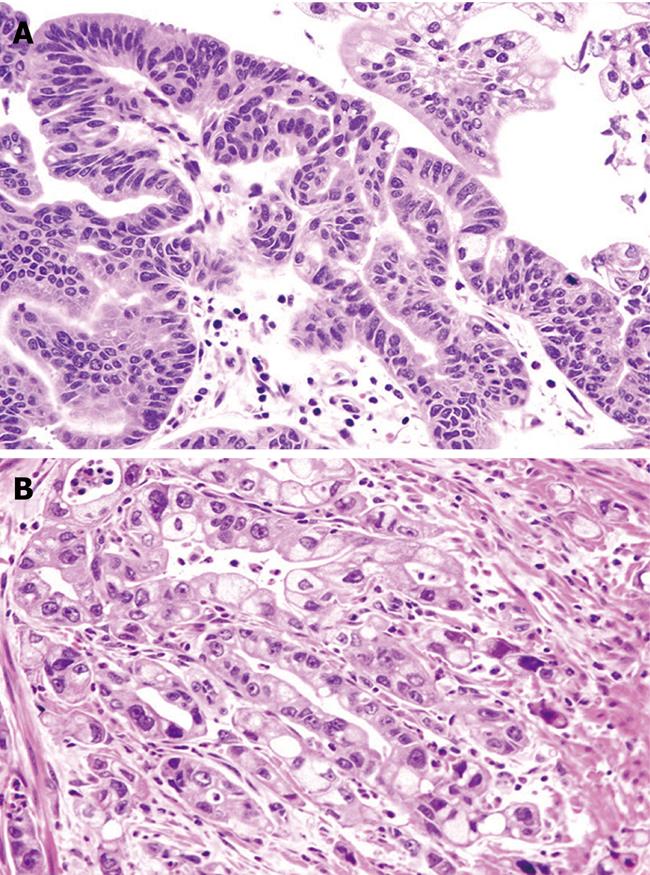
Figure 3 Ampullary tumor, low-grade dysplasia (A) and ampullary adenocarcinoma (B).
Hematoxylin and eosin (HE), original magnification 40 ×.
-
Citation: Manta R, Conigliaro R, Castellani D, Messerotti A, Bertani H, Sabatino G, Vetruccio E, Losi L, Villanacci V, Bassotti G. Linear endoscopic ultrasonography
vs magnetic resonance imaging in ampullary tumors. World J Gastroenterol 2010; 16(44): 5592-5597 - URL: https://www.wjgnet.com/1007-9327/full/v16/i44/5592.htm
- DOI: https://dx.doi.org/10.3748/wjg.v16.i44.5592


