Copyright
©2010 Baishideng Publishing Group Co.
World J Gastroenterol. Nov 28, 2010; 16(44): 5555-5564
Published online Nov 28, 2010. doi: 10.3748/wjg.v16.i44.5555
Published online Nov 28, 2010. doi: 10.3748/wjg.v16.i44.5555
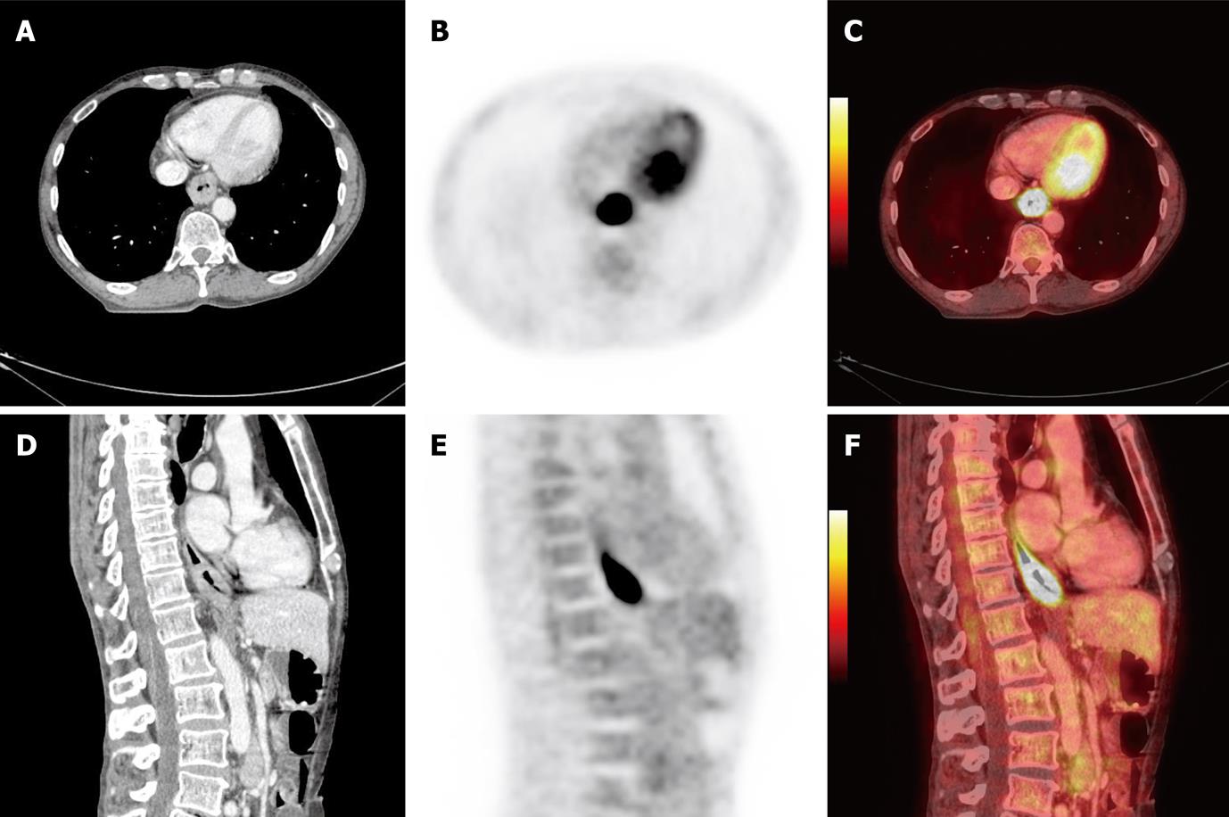
Figure 1 Positron emission tomography/computed tomography images of 61-year-old man with primary squamous cell carcinoma in distal part of the esophagus T3N0M0 prepared in treatment position for radiotherapy planning.
A: Computed tomography (CT), axial slice; B: Positron emission tomography (PET), axial slice; C: PET/CT fusion, axial slice; D: CT, sagital slice; E: PET, sagital slice; F: PET/CT fusion, sagital slice.
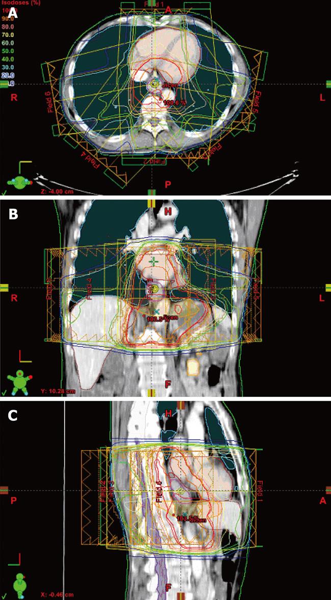
Figure 2 Intensity-modulated radiotherapy plan prepared on a positron emission tomography/computer tomography dataset (Figure 1).
Delineated target volumes and organs at risk, beam arrangement and dose distribution in axial (A), coronal (B), and sagittal slices (C).
- Citation: Vosmik M, Petera J, Sirak I, Hodek M, Paluska P, Dolezal J, Kopacova M. Technological advances in radiotherapy for esophageal cancer. World J Gastroenterol 2010; 16(44): 5555-5564
- URL: https://www.wjgnet.com/1007-9327/full/v16/i44/5555.htm
- DOI: https://dx.doi.org/10.3748/wjg.v16.i44.5555


