Copyright
©2010 Baishideng Publishing Group Co.
World J Gastroenterol. Nov 21, 2010; 16(43): 5502-5509
Published online Nov 21, 2010. doi: 10.3748/wjg.v16.i43.5502
Published online Nov 21, 2010. doi: 10.3748/wjg.v16.i43.5502
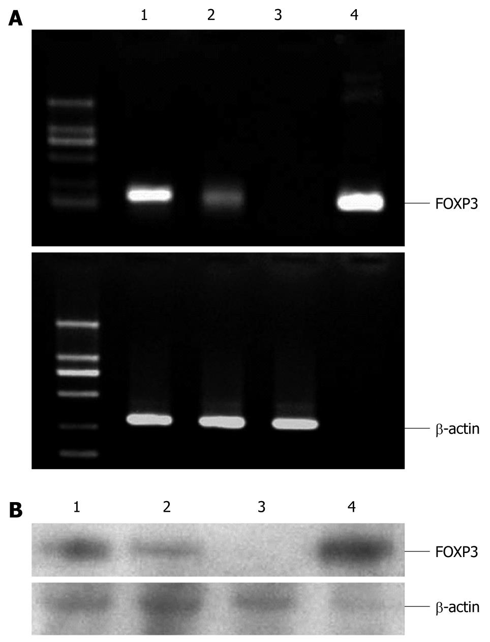
Figure 1 Expression of forkhead box protein 3 in hepatoma cell lines.
A: Expression of forkhead box protein 3 (FOXP3) mRNA is different in hepatoma cell lines by reverse-transcription polymerase chain reaction. β-actin was used to verify the integrity of the template cDNA preparations. 1: SMMC-7721, 2: Hepa-G2, 3: Melanocytes, 4: Foxp3-plasmid; B: Expression of FOXP3 protein was different in hepatoma cell lines by Western blotting. Melanocytes served as a negative control. Foxp3 transiently transferred 293 cells were established as a positive control. 1: SMMC-7721, 2: Hepa-G2, 3: Melanocytes, 4: Foxp3/293 cells.
Figure 2 Flow cytometry of forkhead box protein 3 expression in various hepatoma cell lines.
Melanocytes served as a negative control. The grey underlaid plot represents staining with the isotype, and the white underlaid plot represents staining with anti-human forkhead box protein 3 antibody. FITC: Fluorescein isothiocyanate.
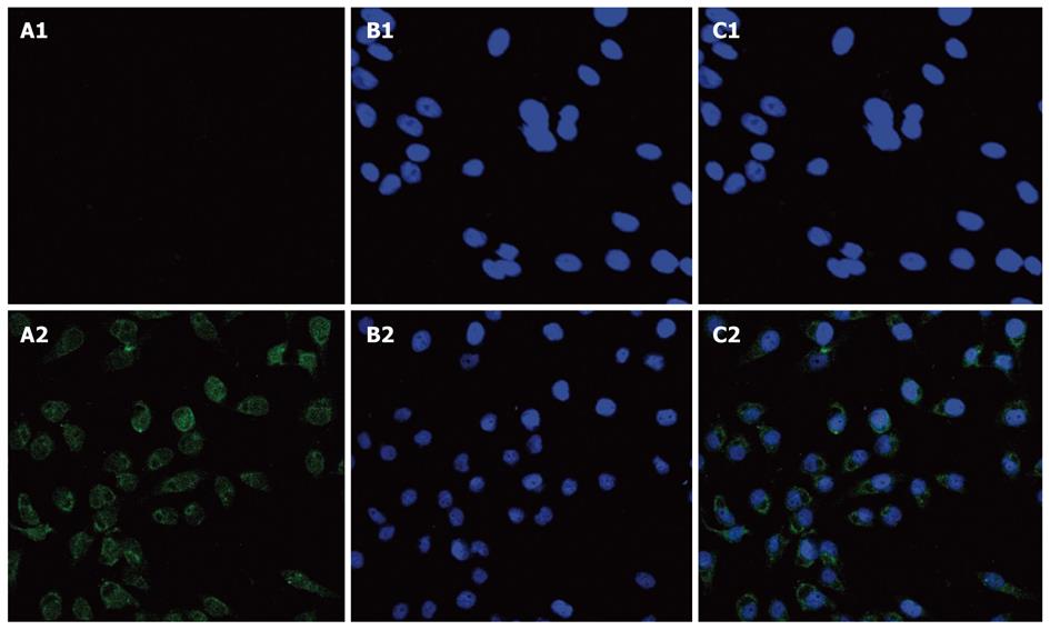
Figure 3 Double-label immunofluorescence analysis of forkhead box protein 3 expression in hepatoma cell line.
A: SMMC-7721 cells express forkhead box protein 3 (FOXP3) in both nuclei and perinuclear cytoplasm (imaged with green fluorescent) (A1: Melanocytes FOXP3-FITC, A2: SMMC-7721 FOXP3-FITC); B: Imaged with diamidino-phenyl-indole (DAPI) to identify the nuclei of SMMC-7721 and melanocytes (B1: Melanocytes DAPI, B2: SMMC-7721 DAPI); C: Image superimposed on a differential interference contrast background confirmed colocalization (C1: Melanocytes merge, C2: SMMC-7721 merge).
Figure 4 Hepatocellular carcinoma immunohistochemical score (× 100).
Forkhead box protein 3 (FOXP3) expression in hepatocellular carcinoma (HCC) by immunohistochemical (IHC). Images represent HCC tissues with IHC scores of 0 (A), 1 (B), 2 (C) and 3 (D). In IHC score 0, many FOXP3-positive lymphocytes are tumor-infiltrating Tregs.
Figure 5 Hepatocellular carcinoma immunohistochemical staining (× 200).
Immunohistochemical (IHC) staining of paraffin embedded hepatocellular carcinoma (HCC) tissues revealed different levels of forkhead box protein 3 (FOXP3) expression in HCC cells (PCH101 antibody, eBioscience). A: Well-differentiated HCC (nuclear FOXP3 staining); B: Moderately-differentiated HCC (cytoplasmic FOXP3 staining); C: Poorly-differentiated HCC (cytoplasmic FOXP3 staining); D: Normal liver tissue (nuclear FOXP3 staining in Tregs).
- Citation: Wang WH, Jiang CL, Yan W, Zhang YH, Yang JT, Zhang C, Yan B, Zhang W, Han W, Wang JZ, Zhang YQ. FOXP3 expression and clinical characteristics of hepatocellular carcinoma. World J Gastroenterol 2010; 16(43): 5502-5509
- URL: https://www.wjgnet.com/1007-9327/full/v16/i43/5502.htm
- DOI: https://dx.doi.org/10.3748/wjg.v16.i43.5502


